Imagine focusing up and down on a miniature cauliflower or closed fist or baseball mitt or tonsil. What would the surface of a raisin look like? A normal nucleus has a relatively round or ovoid shape and its surface is smooth. A dysplastic cell will have humps, bumps, corrugations, crevices, and strange protuberances. These very distinctive abnormalities are the essence of dysplasia, particularly HSIL. This is the very detail that is most often lost in conventional cytology due to the various artifacts of fixation and staining that limit the ability to interpret those conventional smears.
These 3-D nuclear structural abnormalities are to be distinguished from simple, “irregular nuclear outlines” which will often be present as a two-dimensional phenomenon in benign cells on the ThinPrep® Pap Tests. They are two-dimensional in that these “wrinkles” cannot, when the examiner focuses up and down, be traced into the center of the nucleus in the form of crevices, mountains, etc.; they can create deceptive “look-alikes” to the novice.
These 3-D structural abnormalities may not be present in every dysplastic cell on the slide, but they will be obvious in at least some cells somewhere on the slides. Obviously, the ability to see “into” the nucleus of a cell is going to be directly related to the quality of staining. (All ThinPrep® Pap Tests make visualizing cellular changes easier when compared to conventional Pap smears, but over staining or the slightest staleness of reagents will have a direct effect on this the most critical aspect of evaluation.) Also, these 3-D structural defects should be asymmetrical, as opposed to nuclear grooves or simple creases that involve the full breadth of the nucleus occasionally creating a difficult “look-alike”. The presence of these exaggerated nuclear 3-D abnormalities establishes the diagnosis of HSIL.
Reminder: You may click on any slide image
for an enlarged view.
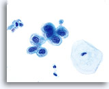
HSIL
Nuclei exhibit asymmetrical 3-D structural abnormalities (difficult to appreciate in 2-dimensional presentation). 60x
HSIL
Nuclei exhibit asymmetrical 3-D structural abnormalities (difficult to appreciate in 2-dimensional presentation). 60x
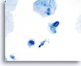
HSIL
Nuclear membranes are markedly irregular and when focused up and down will exhibit humps, bumps, crevices and protuberances. 60x
HSIL
Nuclear membranes are markedly irregular and when focused up and down will exhibit humps, bumps, crevices and protuberances. 60x
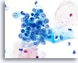
HSIL
Markedly irregular nuclear membranes and 3D structural abnormalities. 60x
HSIL
Markedly irregular nuclear membranes and 3D structural abnormalities. 60x
It is the N/C ratio that is the most reliable indicator of degree (moderate, severe, CIS). With an increasing degree of dysplasia, there is a predictable increasing N/C ratio. This abnormal N/C ratio can be considered a major criterion for the diagnosis of HSIL. However, there are rare exceptions, and ultimately the diagnosis must be made on nuclear changes alone.
Reminder: You may click on any slide image
for an enlarged view.
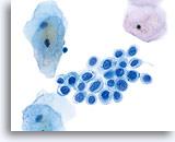
HSIL
N/C ratio is markedly increased and the most reliable indicator of degree of abnormality. 60x
HSIL
N/C ratio is markedly increased and the most reliable indicator of degree of abnormality.
60x
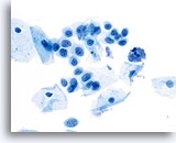
HSIL
With an increasing degree of dysplasia, there is a predictable increasing N/C ratio and this can be considered a major criterion for the diagnosis of HSIL. 60x
HSIL
With an increasing degree of dysplasia, there is a predictable increasing N/C ratio and this can be considered a major criterion for the diagnosis of HSIL. 60x
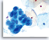
HSIL
Cells of HSIL exhibit an increased N/C ratio. 60x
HSIL
Cells of HSIL exhibit an increased N/C ratio. 60x
There are the essential criteria above (3-D nuclear structural abnormalities and abnormal N/C ratios) necessary to make a diagnosis of HSIL. Then there are other “clues” that are often, or usually found, on a slide demonstrating HSIL. They are as follows:
Reminder: You may click on any slide image
for an enlarged view.
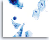
HSIL
Cells often occur singly. Do not expect the streaks of abnormal cells to occur in the strands of mucus seen on the conventional smear. These single cells can be deceptively small. 60x
HSIL
Cells often occur singly. Do not expect the streaks of abnormal cells to occur in the strands of mucus seen on the conventional smear. These single cells can be deceptively small. 60x
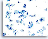
HSIL
These small single cells must be critically examined under high power and are judged by their individual nuclear qualities. 20x
HSIL
These small single cells must be critically examined under high power and are judged by their individual nuclear qualities. 20x
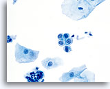
HSIL
Cells often occur in small aggregates and small cobblestone arrangements (often of about 4-7 cells). These groups have a characteristic appearance and are the result of abnormal cohesiveness to be distinguished from “sheets which are probably the simple result of collection device traction. 60x
HSIL
Cells often occur in small aggregates and small cobblestone arrangements (often of about 4-7 cells). These groups have a characteristic appearance and are the result of abnormal cohesiveness to be distinguished from sheets which are probably the simple result of collection device traction. 60x
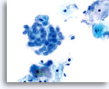
HSIL
Irregularity of nuclear polarity. When in clusters the groups of high-grade nuclei exhibit loss of polarity or a chaotic architecture; they vary in size and shape and have little respect for their neighbors. 60x
HSIL
Irregularity of nuclear polarity. When in clusters the groups of high-grade nuclei exhibit loss of polarity or a chaotic architecture; they vary in size and shape and have little respect for their neighbors. 60x
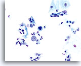
HSIL
Presence of “naked nuclei”. While these are sometimes found in benign (particularly atrophic) smears, they are never to be ignored. This is a very reliable indicator of HSIL. 40x
HSIL
Presence of “naked nuclei”. While these are sometimes found in benign (particularly atrophic) smears, they are never to be ignored. This is a very reliable indicator of HSIL. 40x
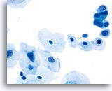
HSIL
The co-presence of more obvious LSIL cells. The concept of “progression” from LSIL to HSIL is somewhat controversial. Although often there will be the larger, more easily recognized cells of LSIL, this is by no means a consistent feature. 60x
HSIL
The co-presence of more obvious LSIL cells. The concept of “progression” from LSIL to HSIL is somewhat controversial. Although often there will be the larger, more easily recognized cells of LSIL, this is by no means a consistent feature. 60x
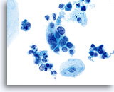
HSIL
Irregularity of nuclear size alone is a much less reliable indicator of dysplasia. 60x
HSIL
Irregularity of nuclear size alone is a much less reliable indicator of dysplasia. 60x
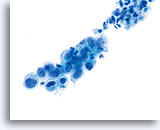
HSIL
Nuclear size variation only has importance when it is observed along with 3-D nuclear structural abnormalities. 60x
HSIL
Nuclear size variation only has importance when it is observed along with 3-D nuclear structural abnormalities. 60x
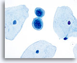
HSIL
Hyperchromasia. Useful when present, however, it is sometimes not present. 60x
HSIL
Hyperchromasia. Useful when present, however, it is sometimes not present. 60x
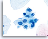
HSIL
Chromatin clumping or granularity. A useful clue when seen in the presence of 3-D deformities and abnormal N/C ratios. By itself however, rarely predictive. 60x
HSIL
Chromatin clumping or granularity. A useful clue when seen in the presence of 3-D deformities and abnormal N/C ratios. By itself however, rarely predictive. 60x
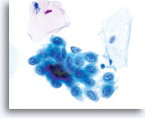
HSIL
Nucleoli generally inconspicuous or absent. Nucleoli may be present when there is a concurrent inflammatory/reactive process or when gland neck involvement is present. 60x
HSIL
Nucleoli generally inconspicuous or absent. Nucleoli may be present when there is a concurrent inflammatory/reactive process or when gland neck involvement is present. 60x
When these “clues” are discovered, there should be a diligent search for the diagnostic cells with undeniable, distinct abnormalities of nuclear structure. Only when these cells are found can a diagnosis of HSIL be made with confidence.
Squamous metaplasia should always be considered a phenomenon independent of dysplasia. When dysplasia occurs in the presence of squamous metaplasia, there must be a “correction” made in the evaluation of N/C ratios. We are theoretically beginning with a cell that, because of squamous metaplasia, already has an abnormal N/C ratio; this therefore becomes less helpful in assigning degree of dysplasia. Do not be “tricked” by N/C ratio in the presence of squamous metaplasia.
Gland neck involvement in HSIL has a distinct appearance on the ThinPrep Pap Test and can be differentiated from lesions of glandular origin. SIL in glands presents predominantly in sheets with increased depth of focus. The cytoplasm is finely vacuolated which initially may give the impression of a glandular process, but on closer inspection these sheets exhibit no glandular differentiation such as basal nuclei, crowded columnar formations, pseudostratification, nor feathered edges or rosettes. These sheets of cells can be deceptively flat, but the nuclei retain the same qualities of SIL that are described above. Because these cells are in sheets and usually are small with no other “clues” of HSIL – only subtle 3-D deformities, they can be the most difficult to identify and evaluate. An important factor in determining whether or not these cells are squamous of glandular in origin is the company they keep. Remember the “like accompanies like” philosophy. Most of the time these cells will be accompanied by definitely dysplastic squamous epithelial cells.
Reminder: You may click on any slide image
for an enlarged view.
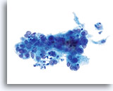
Look-alike
HSIL involving the gland space 40x
Look-alike
HSIL involving the gland space 40x
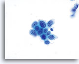
Look-alike
HSIL 60x
Look-alike
HSIL 60x
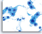
Look-alike
Atrophy 60x
Look-alike
Atrophy 60x
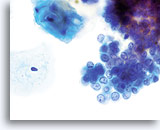
Look-alike
Endometrial cells 60x
Look-alike
Endometrial cells 60x
SUGGESTED READINGS:
- Baker JJ: Conventional and liquid-based cervicovaginal cytology: A comparison study with clinical and histologic follow-up. Diagn Cytopathol 2002;27(3):185-88.
- Bernstein SJ et al: Liquid-based cervical cytologic smear study and conventional Papanicolaou smears: A metaanalysis of prospective studies comparing cytologic diagnosis and sample adequacy. Am J Obstet Gyenecol 2001; 185:308-17.
- Diaz-Rosario LA, Kabawat SE: Performance of a fluid-based, thin-layer Papanicolaou smear method in the clinical setting of an independent laboratory and an outpatient screening population in New England. Arch Pathol Lab Med 1999: 123(9):817-21.
- Selvaggi SM: Cytologic features of high-grade squamous lesions involving endocervical glands on ThinPrep cytology. Diagn Cytopathol 2002;26(3):181-85.
- Weintraub J, Morabia A: Efficacy of a liquid-based thin layer method for cervical cancer screening in a population with a low incidence of cervical cancer. Diagn Cytopathol 200;22(1):52-9.
- Hologic, Inc. ThinPrep® 2000 System Package Insert 2001.


