To further assist in the identification of colloid, a gallery illustrating different morphologic appearances of colloid has been created for use as a reference guide. Additional photographs may be added as cases are collected.
Reminder: You may click on any slide image
for an enlarged view.
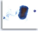
Cyanophilic staining fragment of colloid with frayed edges.
60x
Cyanophilic staining fragment of colloid with frayed edges.
60x
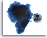
Cyanophilic staining fragment of colloid adjacent to a macrophage containing hemosiderin, red blood cells, and ingested colloid from a cystic lesion.
60x
Cyanophilic staining fragment of colloid adjacent to a macrophage containing hemosiderin, red blood cells, and ingested colloid from a cystic lesion.
60x
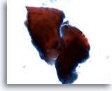
Eosinophilic staining colloid with frayed edges.
60x
Eosinophilic staining colloid with frayed edges.
60x
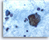
Shiny, refractile colloid in a background of proteinaceous debris.
60x
Shiny, refractile colloid in a background of proteinaceous debris.
60x
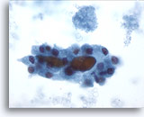
Colloid surrounded by follicular cells exhibiting Hürthle cell change. Lysed blood is in the background.
60x
Colloid surrounded by follicular cells exhibiting Hürthle cell change. Lysed blood is in the background.
60x
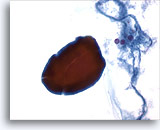
A large, dense fragment of bichromatic staining colloid. Small follicular cells and proteinaceaous debris are in the background.
60x
A large, dense fragment of bichromatic staining colloid. Small follicular cells and proteinaceaous debris are in the background.
60x
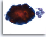
“Cracked” edges of a dense colloid fragment adjacent to benign follicular cells.
60x
“Cracked” edges of a dense colloid fragment adjacent to benign follicular cells.
60x
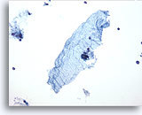
“Tissue-paper” colloid as described by Tulecke and Wang.
20x
“Tissue-paper” colloid as described by Tulecke and Wang.
20x
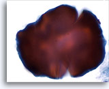
Dense, bichromatic staining colloid.
60x
Dense, bichromatic staining colloid.
60x
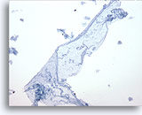
“Tissue-paper” colloid representing watery colloid as described by Tulecke and Wang.
20x
“Tissue-paper” colloid representing watery colloid as described by Tulecke and Wang.
20x
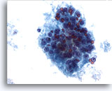
Droplets of colloid of varying size interspersed among benign follicular cells.
40x
Droplets of colloid of varying size interspersed among benign follicular cells.
40x
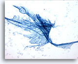
Stringy, shiny colloid as a component of a benign aspirate.
60x
Stringy, shiny colloid as a component of a benign aspirate.
60x
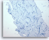
Magnified view of watery colloid.
40x
Magnified view of watery colloid.
40x
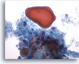
Large fragment of eosinophilic staining colloid with varying size droplets intermixed with follicular cells and debris.
60x
Large fragment of eosinophilic staining colloid with varying size droplets intermixed with follicular cells and debris.
60x
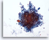
Colloid admixed with benign follicular cells.
20x
Colloid admixed with benign follicular cells.
20x
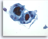
Well-formed colloid is seen within an intact follicle.
60x
Well-formed colloid is seen within an intact follicle.
60x
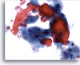
Eosinophilic staining colloid with histiocytes in the background.
40x
Eosinophilic staining colloid with histiocytes in the background.
40x
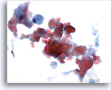
Fragments of colloid are easily recognized at screening power.
20x
Fragments of colloid are easily recognized at screening power.
20x
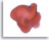
Is this colloid, or something mimicking colloid?
60x
Is this colloid, or something mimicking colloid?
60x
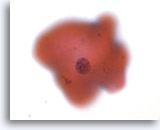
Changing the focal plane reveals that this is not colloid, but an orangeophilic stained, degenerating squamous cell, a colloid “look-alike”. Note the smooth edges and polygonal shape of a typical squamous cell.
60x
Changing the focal plane reveals that this is not colloid, but an orangeophilic stained, degenerating squamous cell, a colloid “look-alike”. Note the smooth edges and polygonal shape of a typical squamous cell.
60x
The following pictures are from malignant thyroid lesions:
Colloid associated with malignant lesions is usually dense, the so called “sticky colloid”.
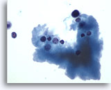
Colloid accompanying a case of medullary carcinoma.
60x
Colloid accompanying a case of medullary carcinoma.
60x
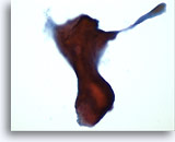
Colloid associated with a case of papillary carcinoma.
40x
Colloid associated with a case of papillary carcinoma.
40x
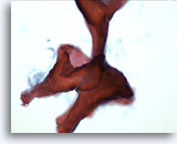
Colloid associated with an additional case of papillary carcinoma.
40x
Colloid associated with an additional case of papillary carcinoma.
40x


