Molecular Biology Text
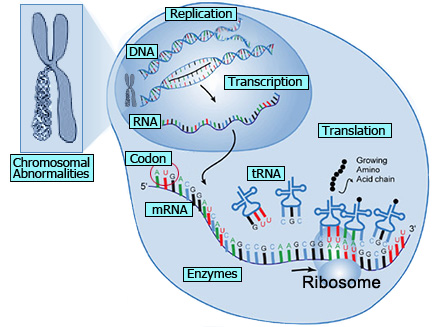
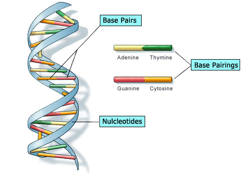
DNA
|
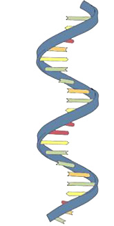
RNA
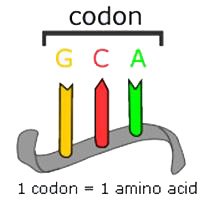
Codon
mRNA
Messenger ribonucleic acid (mRNA) is a molecule of RNA encoding a chemical “blueprint” for a protein product. It is transcribed from the DNA template and carries coding information to the sites of protein synthesis in the ribosomes. In mRNA as in DNA, genetic information is encoded in the sequence of nucleotides arranged into codons consisting of three bases each.
tRNA
Transfer ribonucleic acid (tRNA) is a small RNA molecule that transfers a specific active amino acid to a growing polypeptide chain at the ribosomal site of protein synthesis during translation.
Enzymes
|
Chromosomal Abnormalities
Chromosome abnormalities can be numerical or structural and usually occur as a result of cell
division errors. Fluorescent in situ hybridization utilizes a mixture of multi-colored
fluorescent labeled probes to assess cells for chromosome abnormalities.
Anueploidy is defined as the occurrence of one or more extra or missing chromosomes
leading to an unbalanced chromosome complement, or, any chromosome number that is not an exact multiple
of the haploid number. Abnormal chromosome number is one of the most common features of cancer
cells and is caused by many mechanisms.
Nondisjunction is the failure of chromosome pairs to separate properly during anaphase, resulting
in a cell with chromosome imbalance. This failure to separate is frequently caused by spindle
checkpoint defects. Spindle, or mitotic, checkpoints are mechanisms that control the proper cell
division in eukaryotic cells. To achieve proper cell division, the two kinetochores on the
sister chromatids must be attached to opposite spindle poles. Spindle checkpoints assess the cell for
DNA damage and will delay division or target the cell for destruction if repairs are not
accurately completed. A weakened or damaged spindle checkpoint would generate daughter cells either
lacking a chromosome copy or possessing too many copies.
Aneuploidy is frequently found in bladder cancer. Bladder cancer is the fourth most common
cancer in men and the seventh most common cancer in women in the United States. Bladder cancer
recurrence rate is 80%, making it a chronic illness. Diagnosis of low grade and recurrent bladder
cancer has been difficult due to the low sensitivity of urine cytology and the cost and
invasiveness of cystoscopy . A new FISH assay has been developed that tests for aneuploidy of the
chromosomes 3, 7 and 17 frequently associated with bladder cancer.
Deletion is the loss of a part of the chromosome or DNA sequences. Loss of genetic material can
range from a single nucleotide base pair to an entire piece of chromosome and can be caused by
losses from translocation , unequal crossing over, chromosomal crossovers within a
chromosomal inversion and breaking without rejoining. FISH can be used to detect the loss of
the 9p21 locus found in more than 60% of bladder cancers.
Duplication is the production of multiple copies of a region of DNA that contains a gene; it may
occur as an error in a retrotransposition event, a homologous recombination, or duplication of an entire
chromosome. FISH is the gold standard for detecting amplification of the HER2/neu ( Human
Epidermal growth factor Receptor 2) gene or its protein product. Overexpression of this receptor
is found in 15 to 20 percent of breast cancers and also occurs in ovarian cancer, stomach cancer, and
serous endometrial carcinoma.
Chromosomal translocation is an abnormality caused when a portion of one chromosome is
transferred to a nonhomologous chromosome . Cancer, infertility and a minority of Down’s
syndrome cases can be attributed to this event. Chromosomal inversion occurs when a piece of
chromosome breaks off and reattaches itself in the reverse direction. Carriers of this
defect usually do not experience any abnormalities if the chromosome rearrangement does not result in
missing or additional genetic information.
References:
- Text – Alberts, Bruce, et al. (2002) The Molecular Biology of the Cell (4th edition). Garland Science
- Text – Bruns, David E, et al. (2007) Fundamentals of Molecular Diagnostics. Saunders
- Text – Interactive Medical (2009). Molecular Diagnostic Testing: Principles and Practice. CE and CME program. www.hpvinstitute.com.
- Image – http://ghr.nlm.nih.gov/handbook/illustrations/dnastructure.jpg (DNA strand)
- Image – http://images2.clinicaltools.com/images/gene/codon.jpg (RNA strand)
- Image – http://www.obgynacademy.com/basicsciences/fetology/genetics/images/codon_GCA.gif
- Image – http://www.theodora.com/genetics/glossary_t.html (transcription/translation)
Click here to return to the Molecular Biology section