Fluorescence In Situ Hybridization Lab
Move your mouse over any label for a detailed description.
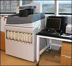
There are small bench top instruments (16″ x 9″) that are “semi-automatic” and can handle 12 slides at a time. Also available are instruments which will completely automate the process of FISH including the baking, deparaffinization (if necessary), cell conditioning and staining.
Click
 again to see an image of this type of instrument.
A fluorescence microscope is necessary to analyze FISH specimens. There have been many improvements to microscopic optics and filter technology in the past two decades.
Visual analysis is a labor- intensive and often subjective procedure without the use of advanced fluorescence imaging. Image analysis software is widely available from many manufacturers.
References
Text – http://jcs.biologists.org/cgi/content/full/116/14/2833
Text – Lu, Yong-Jie, et al. An accelerated in situ hybridization procedure using microwave irradiation. Chromosome Research Vol 5, 1997. Rapid Science Publishers.
Text – http://osiris.rutgers.edu/~smm/in_situ_hybridization.htm
Text – http://www.intavis.com/en/In_Situ_Detection/index.php
Text – http://www.microscopyu.com/articles/fluorescence/insitu/brennerinsitu.html
Text – Interactive Medical (2009). Molecular Diagnostic Testing: Principles and Practice. CE and CME program. www.hpvinstitute.com.
Images – http://uk.precisionnews.com/article_Transfert-libre-a_1861.html
Images – http://www.dako.com
Images – http://www.umanitoba.ca/institutes/manitoba_institute_cell_biology/GCCRD/imaging_systemsNew.htm
Images – http://www.unr.edu/inbre/cores/histology/Instrumentation.asp
Click here to close this window.
Fluorescence in situ hybridization (FISH) is a gene expression technique which localizes specific nucleic acid targets in fixed tissue and cells. Various types of cytogenetic alterations including aneusomy, duplication, amplification, deletion, and translocation that occur with neoplasia in exfoliative and aspiration cytology specimens can be detected using FISH. Although the technique is about 20 years old, automation is fairly recent. In situ detection protocols can be time consuming; taking several days with labor intensive repeated washing and incubation steps. Microwaves can be used in rapid FISH protocols to improve process times. Automation offers an increase in throughput and a reduction of human error by the accurate control of temperature, correct pipetting volume and proper incubation time.
Click here to learn more about fluorescence in situ hybridization methods.
Return to Instrumentation Section
- Text – http://jcs.biologists.org/cgi/content/full/116/14/2833
- Text – Lu, Yong-Jie, et al. An accelerated in situ hybridization procedure using microwave irradiation. Chromosome Research Vol 5, 1997. Rapid Science Publishers.
- Text – http://osiris.rutgers.edu/~smm/in_situ_hybridization.htm
- Text – http://www.intavis.com/en/In_Situ_Detection/index.php
- Text – http://www.microscopyu.com/articles/fluorescence/insitu/brennerinsitu.html
- Text – Interactive Medical (2009). Molecular Diagnostic Testing: Principles and Practice. CE and CME program. www.hpvinstitute.com.
- Images – http://uk.precisionnews.com/article_Transfert-libre-a_1861.html
- Images – http://www.dako.com
- Images – http://www.umanitoba.ca/institutes/manitoba_institute_cell_biology/GCCRD/imaging_systemsNew.htm
- Images – http://www.unr.edu/inbre/cores/histology/Instrumentation.asp