

Case Presentation
Case Presentation – August 2026
Metastatic Papillary Thyroid Carcinoma
Written by: Mia Schloneger, Student, Cleveland Clinic School of Health Professions Cytology Program, Cleveland, Ohio
Patient Age: 59-year-old female
Specimen Type: 3p (retrotracheal) lymph node EBUS TBFNA: Modified Romanowsky-stained and Pap-stained smears, Pap-stained ThinPrep® Non-Gyn slide, Hematoxylin and Eosin (H&E)-stained Cellient® cell block. Corresponding surgical pathology.
Patient History: Papillary thyroid cancer (PTC) followed by a thyroidectomy, isthmusectomy and neck dissections (left lateral and central neck dissection and left modified radical neck dissection), metastatic PTC to lymph nodes in the central neck, innumerable small lung metastases, and radioactive Iodine ablation to the right thyroid.
Cytologic Diagnosis: Positive for malignant cells. Metastatic papillary thyroid carcinoma.
Biopsy / Pathologic Diagnosis: 3p lymph node core biopsy, surgical specimen: H&E-stained slides. Core biopsy was positive for papillary thyroid carcinoma, classic (conventional) subtype. IHC stains were positive for TTF-1, PAX-8, Thyroglobulin, and BRAF V600E.
Case provided by: Cleveland Clinic, Cleveland, Ohio
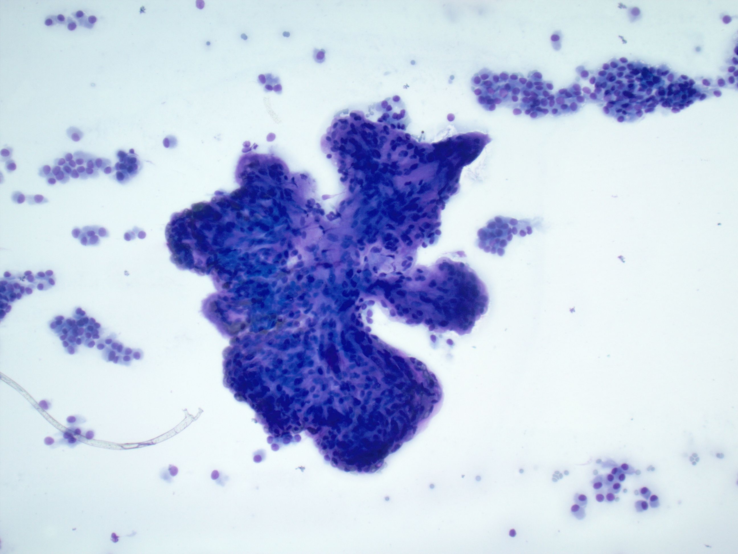
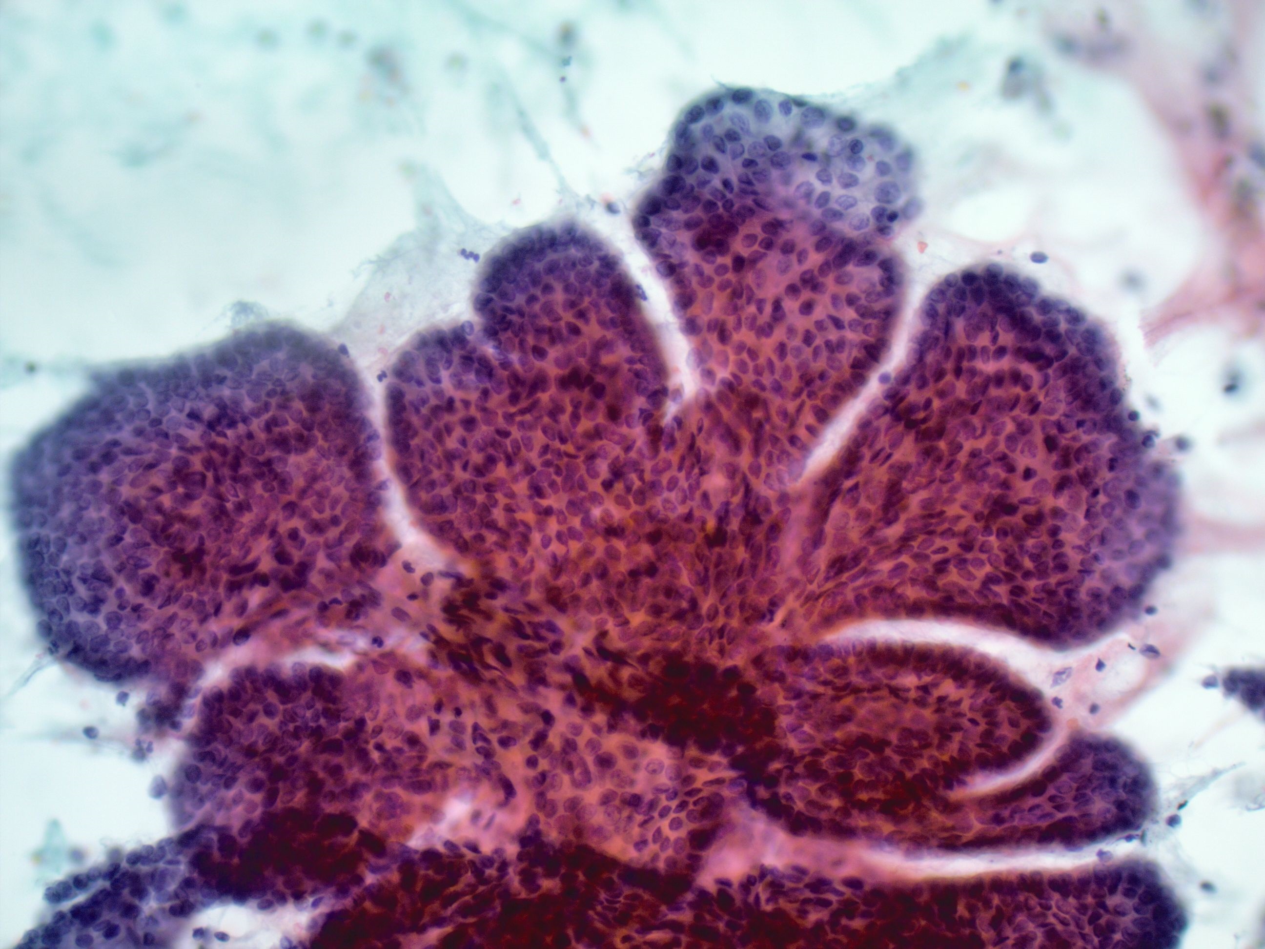
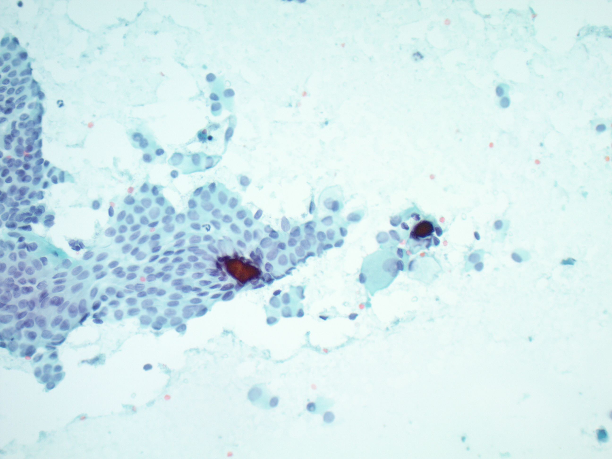
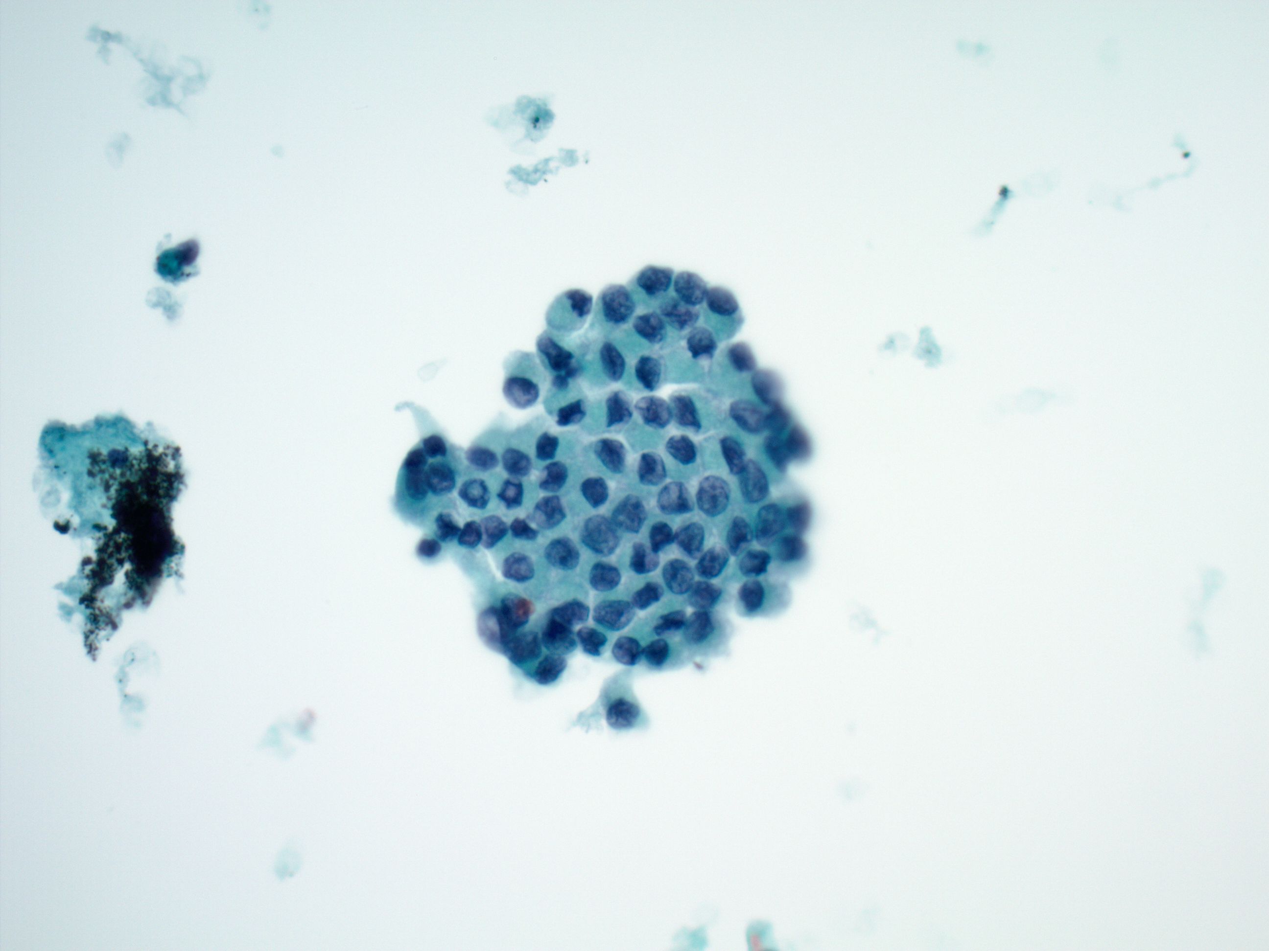
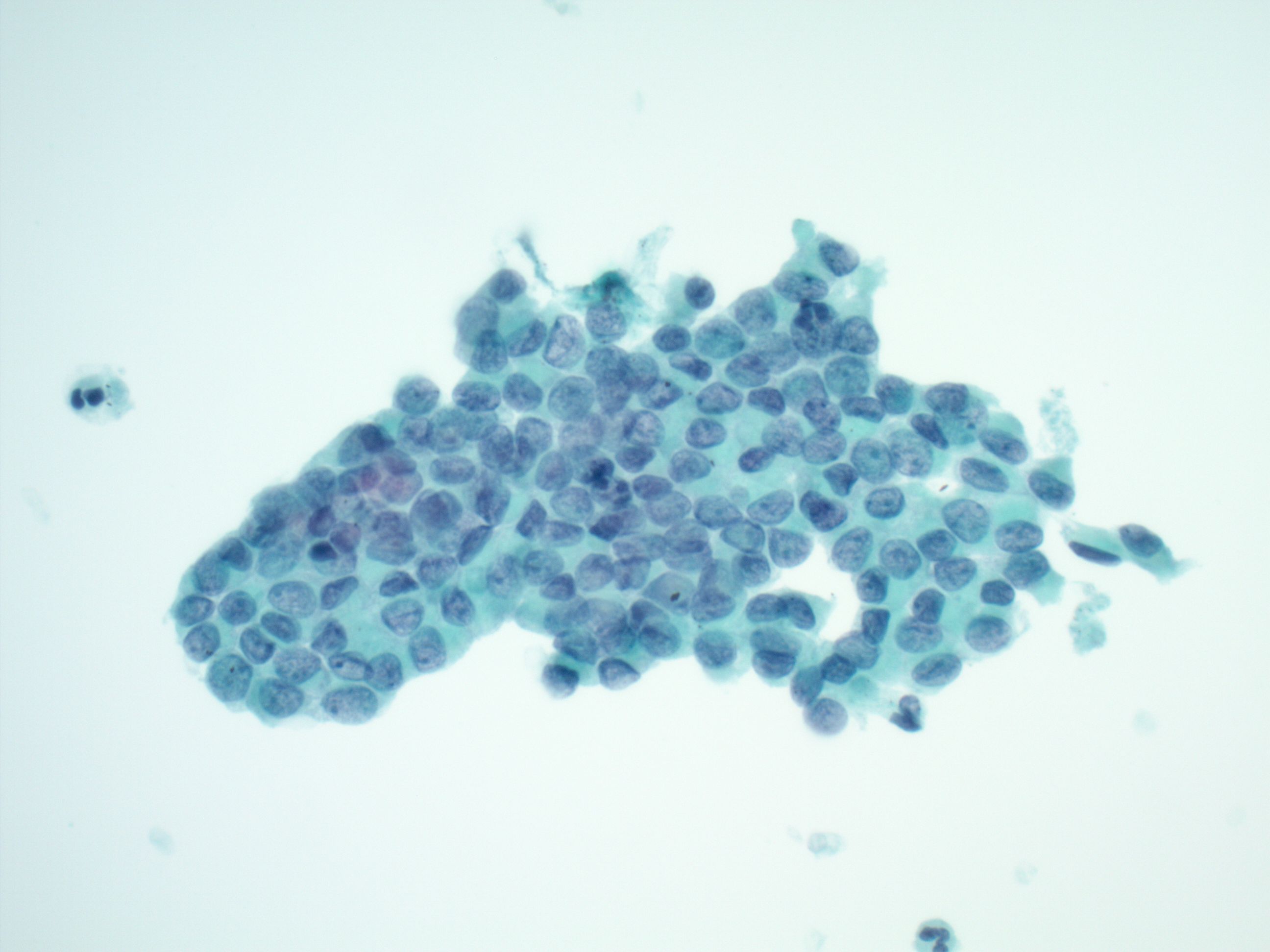
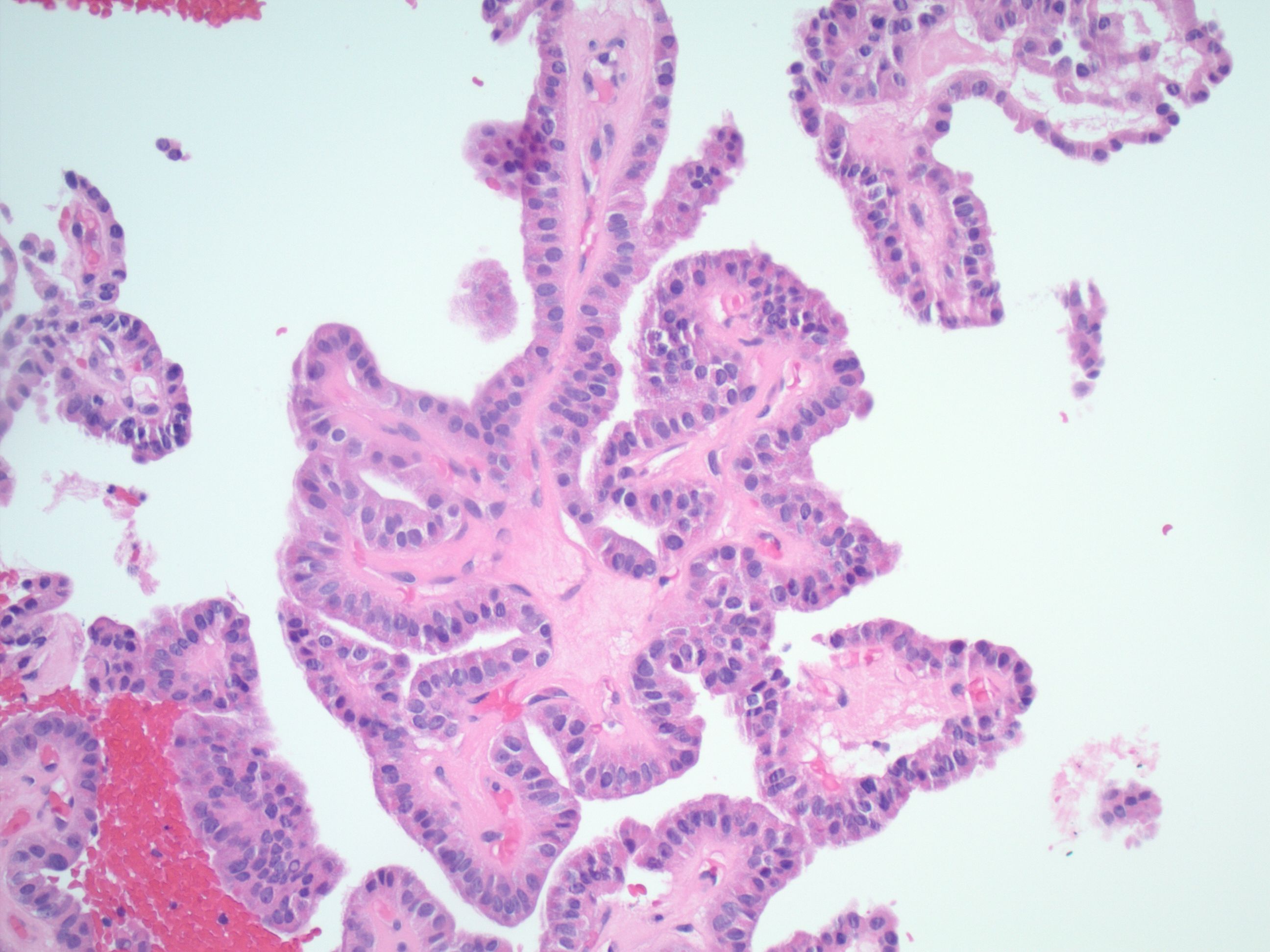
Metastatic Papillary Thyroid Carcinoma
Etiology:
Thyroid Cancer is the 13th most common cancer diagnosis in the U.S. with more than 45,000 new cases estimated in 2026. In recent years, the number of cases diagnosed has been on the rise due to frequent findings on imaging scans.1,2 Papillary thyroid carcinoma (PTC) is the most common histologic type, accounting for over 85% of thyroid carcinomas.3 Although seen in men, PTC is 4 times more common in women, often thought to be due to higher levels of estrogen production.2,4 PTC is more common in younger patients with a mean age at diagnosis of 51 years, but most presenting before the age of 40.5 A leading factor for the early onset diagnosis is high exposure to radiation during childhood, greatly increasing the risk of developing PTC for both men and women.4,6 Increased iodine intake is another factor for increased risk, as excess iodine intake causes an imbalance in thyroid hormones and alters thyroid function.7 Although recognizing these factors is important, often patients who develop PTC will present with no predisposing risks.5 This variability in risk assessment can make a PTC diagnosis challenging before metastatic spread occurs. Metastases to regional lymph nodes are common in PTC, as in many other malignancies. Up to 20% of patients will have metastasis involving the lymph nodes at first diagnosis.5,6 Metastasis to uncommon lymph node regions, such as the retrotracheal nodes is rare, which is why this area is not routinely sampled.8 Although less common, PTC can also metastasize to more distant sites including the lungs.4,6
Clinical Features:
PTC may present with a variety of clinical features. Often it appears as a palpable thyroid nodule, which may cause dysphagia, voice changes, or pain. Typically, these nodules are slow-growing, and the patient may be asymptomatic altogether.4,5 Metastatic PTC to the lymph nodes and lungs, as seen in this patient, may cause additional symptoms. Symptoms such as lymphadenopathy, shortness of breath, or chest pain are common.5
Treatment and Prognosis:
Metastatic cancers often have poor prognoses, and they are more difficult to treat. However, the prognosis for PTC remains very good, even when metastasized. The 5-year survival rate for patients with regional metastasis of PTC is 99% and survival remains high for those with distant metastases, including spread to the lung.9 Involved lymph nodes usually have no impact on prognosis but often will increase the chance of reoccurrence.5,10 Genetic drivers also are a factor affecting the prognosis and development of PTC. Patients with BRAF V600E mutation have been associated with increased mortality and recurrence when compared to those without, making treatment and management more complex.2,6 Treatment of PTC is often a lobectomy or total thyroidectomy dependent on clinical factors of the tumor and the patient. This may be followed by radioiodine therapy especially for targeting metastases.3,6 A lymphadenectomy can also be performed to remove any metastatic involvement of regional nodes.10
Cytology:
Metastatic PTC often shows cytologic features similar to those of the primary tumor. Distinct nuclear features that are important for this diagnosis are enlarged and crowded nuclei, nuclear grooving, powdery chromatin, and prominent nucleoli. Architecturally, these cells can be arranged in papillary structures with fibrovascular cores or as flat sheets. Some additional features that support this diagnosis, but are not exclusive to PTC, include psammoma bodies and intranuclear pseudoinclusions.3,6 These features were clearly demonstrated in this case. Immunohistochemical (IHC) stains are often helpful in distinguishing metastasis from a new primary cancer. Thyroglobulin, PAX-8, and TTF-1 all stain positive in PTC and are important in confirming this diagnosis. 3,6
Differential Diagnosis:
Assessment of differential diagnoses is key for ensuring all possibilities are considered for the best diagnosis. Since this patient had lung nodules, it is important to first rule out metastatic lung cancer. Metastatic lung adenocarcinoma can display papillary architecture with fibrovascular cores, similar to PTC. However, adenocarcinomas will have nuclei with coarse chromatin and prominent nucleoli. Since both lung adenocarcinoma and PTC stain positive for TTF-1, a positive Napsin-A result can help favor a lung adenocarcinoma diagnosis in the appropriate clinical context.3,11 PAX-8 is virtually never positive in primary lung adenocarcinoma, therefore positivity for PAX-8 would favor thyroid origin.12
Another lung primary that could be considered in this case is a carcinoid tumor. Carcinoid tumors are generally slow-growing and usually will not metastasize; when they do, they can present similar features to PTC. Loose clusters and single cells may aggregate around transgressing vessels, mimicking a papillary formation. Carcinoid tumors present with “salt-and-pepper” chromatin, in contrast to the pale chromatin seen in PTC. IHC stains synaptophysin and chromogranin are useful in this differential.11
Morphologically, ovarian adenocarcinoma can also display papillary structures with fibrovascular cores, similar to those seen in PTC. Psammoma bodies, which were prominent in this case of PTC, are also a feature frequently found in ovarian adenocarcinoma. Since PTC and ovarian adenocarcinoma both stain positive for PAX-8, markers such as ER/PR can help exclude ovarian adenocarcinoma as the source.11
Although IHC can be helpful in refining the differential diagnosis, clinical context and morphology are also crucial factors in arriving at the correct diagnosis. In this case of a patient with a history of PTC, innumerable small pulmonary nodules and no history of a pelvic mass, the morphologic features (in combination with positivity for TTF-1, PAX-8, thyroglobulin, and BRAF V600E) are consistent with metastatic PTC.
References:
- American Cancer Society. Key Statistics for Thyroid Cancer. Published January 13, 2026. Accessed February 16, https://www.cancer.org/cancer/types/thyroid-cancer/about/key-statistics.html
- Kitahara CM, Schneider AB. Epidemiology of Thyroid Cancer. Cancer Epidemiology, Biomarkers & Prevention. 2022;31(7):1284-1297. doi:https://doi.org/10.1158/1055-9965.epi-21-1440
- Ali SZ, VanderLaan The Bethesda System for Reporting Thyroid Cytology. Springer Nature; 2023: 135-147, 171-172, 232-233.
- Mayo Thyroid cancer – Symptoms and causes. Mayo Clinic. Published January 5, 2024. Accessed February 16, 2026. https://www.mayoclinic.org/diseases-conditions/thyroid-cancer/symptoms-causes/syc-20354161
- Columbia Thyroid Center. Guide to Papillary Thyroid Columbiasurgery.org. Published 2025. Accessed February 20, 2026. https://columbiasurgery.org/conditions-and-treatments/papillary-thyroid-cancer#signs-and-symptoms.
- Faten Limaiem, Rehman A, Mazzoni Papillary Thyroid Carcinoma. StatPearls [Internet]. Published March 13, 2024. Accessed February 21, 2026. https://www.ncbi.nlm.nih.gov/books/NBK536943
- Kim K, Cho SW, Park YJ, Lee KE, Lee DW, Park Association between Iodine Intake, Thyroid Function, and Papillary Thyroid Cancer: A Case-Control Study. Endocrinology and Metabolism. 2021;36(4):790-799. doi:https://doi.org/10.3803/enm.2021.103
- Shao K, Gao SG, Xue Q, et Clinical analysis of mediastinal lymph node dissection through sternotomy approach in the treatment of papillary thyroid carcinoma with mediastinal lymph node metastasis. Zhonghua yi xue za zhi. 2020;100(24):1866-1871. doi:https://doi.org/10.3760/cma.j.cn112137-20200428-01353
- American Cancer Survival Rates for Thyroid Cancer. www.cancer.org. Published June 30, 2025. Accessed February 21, 2026. https://www.cancer.org/cancer/types/thyroid-cancer/detection-diagnosis-staging/survival-rates.html
- Maksimovic S, Branislava Jakovljevic, Zdenka Lymph Node Metastases Papillary Thyroid Carcinoma and their Importance in Recurrence of Disease. Medical Archives. 2018;72(2):108-108. doi:https://doi.org/10.5455/medarh.2018.72.108-111.
- Cibas ES, Ducatman Cytology : Diagnostic Principles and Clinical Correlates. 5th ed. Elsevier; 2020: 88-90, 94-96, 527-528.
- McHugh KE, Arrossi AV, Farver CF, Mukhopadhyay S. Does Strong and Diffuse PAX-8 Positivity Occur in Primary Lung Carcinoma? An Immunohistochemical Study of 418 Cases and Review of the Literature. Appl Immunohistochem Mol Morphol. 2019;27(2):140-146. doi:10.1097/PAI.0000000000000549