Case Presentation
Case Presentation – August 2022
Alveolar Rhabdomyosarcoma
Written by: Harley Moser, Student, Cleveland Clinic School of Cytotechnology, Cleveland Ohio
Patient Age: 25-year-old, Female
Specimen Type: FNA of 4R lymph node, Traditional smears with Diff Quik and pap stain, ThinPrep® Non-Gyn, Cell Block
Patient History: Prior diagnosis of alveolar rhabdomyosarcoma with PAX3-FOXO1 gene fusion
Cytologic Diagnosis: Positive for malignant cells derived from metastatic rhabdomyosarcoma
Biopsy / Pathologic Diagnosis: Metastatic alveolar rhabdomyosarcoma
Case provided by: Cleveland Clinic
Alveolar Rhabdomyosarcoma
Etiology:
Rhabdomyosarcoma (RMS) is derived from rhabdomyoblasts with skeletal muscle differentiation.1 RMS is divided into four subgroups – alveolar rhabdomyosarcoma (ARMS), embryonal rhabdomyosarcoma (ERMS), pleomorphic rhabdomyosarcoma, and spindle cell rhabdomyosarcoma. The most frequent subtype is ERMS, which is broken down into two variants – botryoides and anaplastic. ARMS is the second major subtype with two variants – solid and anaplastic. Additional classification of RMS is molecular identification for “fusion positive” and “fusion negative” between the two proteins, PAX3 or PAX7 and FOXO1.1,2 Currently, the cause of this malignancy is unknown, but some research suggests risk factors include genetic syndromes (Li-Fraumeni, Gardner, and Beckwith-Wiedemann), prenatal X-ray exposure, maternal drug use, fertility medications, premature birth, first-degree relative with RMS, and allergies.3
Clinical Features:
Rhabdomyosarcoma most commonly affects children and adolescents, and rarely occurs in adults.4 Although RMS is predominantly found on the head and neck, ARMS is more frequently located on the extremities. ARMS patients will usually present with a painless mass on the primary site leading to possible joint pain, redness, and discomfort.1 Once ARMS diagnosis is made, metastases to the bones, lungs, and lymph nodes are commonly found. In our case, the patient presented with an enlarging perianal mass with left iliac metastases.
Treatment and Prognosis:
Pediatric RMS has a good prognosis with a >70% five-year survival rate.1 However, RMS in adults is very aggressive with a poor prognosis. The alveolar subtype is usually found with metastasis or at advanced stages having a five-year survival rate of 5-15%.5 The best course of treatment is surgical resection with negative margins, chemotherapy, and radiotherapy. Patients with metastasis have a better prognosis if radiotherapy is started right after surgical removal because of the high rate of metastasis. Chemotherapy is based on the subtype and whether it is fusion-positive or fusion-negative.
Cytology:
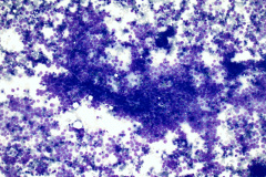
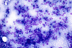
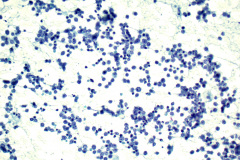
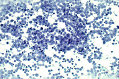
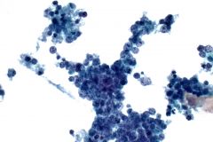
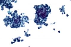
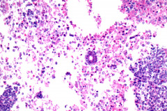
Alveolar RMS morphologically resembles a small round blue cell tumor with irregular alveolar structures seen in histology.1 The tumor cells are uniformly round or polygonal but can appear as tadpole or ribbon-like at times. The cytoplasm is granular, eosinophilic, and glycogen vacuoles may be seen. Nuclei are enlarged with multiple prominent nucleoli and coarse chromatin. Necrosis is noteworthy with some rhabdomyoblasts present in the background.6, 7 Benign rhabdomyoblast criteria is based on their myogenic differentiation – early, intermediate, or late rhabdomyoblasts. Early rhabdomyoblasts are identical to the small round blue tumor cells, while intermediate and late rhabdomyoblasts have abundant cytoplasm and irregular nuclei. Our case presented with all the criteria listed, but tadpole cells were not seen. The slides were hypercellular with marked necrosis. Alveolar rhabdomyosarcoma stains positive with the following IHC markers – myogenin, desmid, MyoD1, CAM 5.2, and chromogranin.8
Differential Diagnosis:
Neuroblastoma is the most common extracranial childhood malignancy.6 The cytology is a classic small round blue cell tumor like ARMS. Homer-Wright rosettes containing neutrophils will be present, but they are not a prominent feature. Nuclei are small, round, and hyperchromatic with little to no eosinophilic cytoplasm. The IHC stains that are positive for neuroblastoma are NSE, CD57, CD56, synaptophysin, and chromogranin.
Lymphoma/Leukemia is a bone tumor that can occur at any age but is more commonly found in adults.7 The cytologic pattern is single cells with scant cytoplasm. A frequent characteristic is nuclei with clefts in the nuclear membrane and prominent nucleoli. Positive IHC stains for hematopoietic neoplasms are CD45, CD5, CD30, CD19, CD3, and CD10.
Ewing Sarcoma is a common high grade bone sarcoma in children and adolescents. Cytologically, the cells are predominantly single in a “tigroid” background.7 There is a high nuclear-to-cytoplasmic ratio with the cells being around two to three times the size of a lymphocyte. A prominent feature is nuclear molding, which is not seen in alveolar rhabdomyosarcoma. This sarcoma stains positive with the following IHC stains – CD99, NKX2.2, Vimentin, FLI1, and ERG.10
Poorly Differentiated Synovial Sarcoma is an aggressive neoplasm that commonly affects joints in an extremity.6 Although the cytology appears as a small round blue cell tumor, there are also cohesive, oval to spindle cells present. Unlike the other differentials listed, this sarcoma has a vascular hemangiopericytoma-like pattern with pseudorosettes.7 Common stains that are positive are TLE1, CK7, CK19, EMA, CD56, and BCL2.11
References:
- Skapek SX, Ferrari A, Gupta AA, Lupo PJ, Butler E, Shipley J, Barr FG, Hawkins DS. Rhabdomyosarcoma. Nat Rev Dis Primers. 2019 Jan 7;5(1):1
- Dziuba I, Kurzawa P, Dopierała M, Larque AB, Januszkiewicz-Lewandowska D. Rhabdomyosarcoma in children – current pathologic and molecular classification. Pol J Pathol. 2018;69(1):20-32.
- Gartrell J, Pappo A. Recent advances in understanding and managing pediatric rhabdomyosarcoma. F1000Res. 2020 Jul 8; 9:F1000 Faculty Rev-685.
- He W, Jin Y, Zhou X, Zhou K, Zhu R. Alveolar rhabdomyosarcoma of the vulva in an adult: a case report and literature review. J Int Med Res. 2020 Mar;48(3)
- Dondapati M, Reyes JVM, Ahmad S, Stern AS, Lieber JJ. Rare Adult Subtype of Rhabdomyosarcoma, a Common Childhood Soft Tissue Carcinoma. J Investing Med High Impact Case Rep. 2021 Jan-Dec;9:23247096211042236
- DeMay R. The Art and Science of Cytopathology, Second Edition. 2012. American Society for Clinical Pathology.
- Cibas E. S., and Ducatman B. S. Cytology: Diagnostic Principles and Clinical Correlates: Fifth Edition. 2021. Elsevier Inc.
- Ozer E. Alveolar rhabdomyosarcoma. PathologyOutlines.com website. https://www.pathologyoutlines.com/topic/softtissuealvrhabdo.html.
- Parham DM, Barr FG. Classification of rhabdomyosarcoma and its molecular basis. Adv Anat Pathol. 2013 Nov;20(6):387-97
- Warmke L. Ewing sarcoma. PathologyOutlines.com website. https://www.pathologyoutlines.com/topic/boneewing.html.
- Obeidin F, Alexiev BA. Synovial sarcoma. PathologyOutlines.com website. https://www.pathologyoutlines.com/topic/softtissuesynovialsarc.html.

