Case Presentation
Case Presentation – August 2021
Basaloid Squamous Cell Carcinoma in the Lungs
Written by: Loc Pham, Student, Cleveland Clinic School of Cytotechnology, Cleveland, Ohio
Patient Age: 81-year-old female
Specimen Type: Bronchial FNA of the 4L lymph node, Diff Quik Smear, ThinPrep® Non-Gyn
Patient History: Former smoker of 42 years. No history of dysplasia.
Cytologic Diagnosis: Positive for malignant cells. Poorly differentiated carcinoma, most consistent with non-keratinizing squamous cell carcinoma.
Case provided by: Dr. Fatimah Hamadeh, Cleveland Clinic Cytopathologist
Basaloid Squamous Cell Carcinoma in the Lungs
Etiology:
The World Health Organization defined basaloid squamous cell carcinoma (BSCC) as an aggressive, high-grade, variant of squamous cell carcinoma (SCC).1 The tumor has both basaloid and squamous features. It is commonly found in the head and neck region and can be found less commonly at the trachea.2 Most patients reported having smoked for a long time before the discovery of the tumor.3 In this case, the patient smoked for 42 years and the tumor was found in the patient’s lungs, lymph nodes, and a later PET scan revealed multiple metastases to her liver, right adrenal, left kidney, mesentery, chest wall, rectum, and mediastinum.
Clinical features:
Similar to conventional SCC, BSCC can present as a painless irregular mass that may or may not be ulcerative. While conventional SCC is common in the lungs, BSCC is more common in the head and neck area and reported clinical symptoms include burning sensation of the tongue area, swelling of the neck, etc.4,5 In this case, the patient presented with dyspnea, exertional shortness of breath, wheezing episodes and loss of appetite without weight loss. The thoracic imaging revealed a 14.5 cm mass in the infrahilar region of the right lower lobe of the right lung, with thoracic lymphadenopathy concerning for bronchogenic neoplasm. A 1.4 cm nodule was also found in the right adrenal gland nodule as well as a 1.8 cm dense structure in the left kidney.
Treatment and Prognosis:
BSCC is an aggressive variant of SCC and frequently recurs locally with regional and distant metastases. Most are usually diagnosed at advanced clinical stages and thus prognosis is unfavorable.6 In this case, PET scan revealed that the tumor has metastasized to the patient’s liver, right adrenal, left kidney, mesentery, chest wall, rectum, and mediastinum. Thus, her treatment is palliative chemoimmunotherapy with carboplatin, paclitaxel and pembrolizumab iv (with prochlorperazine as needed).
Cytology:
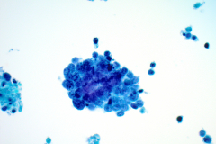
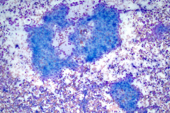
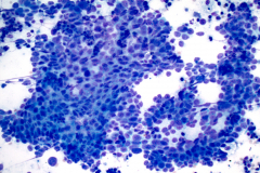
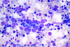
BSCC reportedly display very high nuclear/cytoplasmic ratio with coarse chromatin, inconspicuous to prominent nucleoli and nuclear pleomorphism. The nuclei also mold, and the cells tend to arrange in cohesive clusters, with necrosis in the background.6 This case demonstrates all these criteria (see figures 1-4).
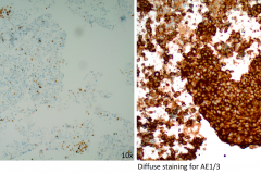
The cells of BSCC are positive for SCC markers p63 and p40.8, 9 Cytokeratin markers such as CK1, CK7 and CK14 are also positive in BSCC.10 In this case, AE1/3 was used to confirm epithelial differentiation11 along with p40 to confirm squamous differentiation (see figure 5). BSCC cells are negative for neuroendocrine markers synaptophysin and chromogranin, adenocarcinoma markers, and lymphoid markers.11, 12
Differential Diagnosis:
- Small cell carcinoma is a common tumor found in the lungs of patients who had extensive history of smoking. The cells display high N/C ratio with neuroendocrine chromatin. The nuclei tend to mold and generally do not show prominent nucleoli. The cells can form loose clusters while the background contains a large amount of necrotic materials. Small cell carcinoma cells stain positive for synaptophysin, chromogranin, TTF-1, and CD56. They are negative for p63, p40 and CD45.11, 12
- Lymphoma can also be found in the lungs due to the extensive network of lymph nodes in the area. The cells also have high N/C ratio with coarse chromatin that can display prominent nucleoli and are dishesive. A variant of lymphoma, Hodgkin lymphoma, can also display nuclear pleomorphism and can have loose clusters at times. Lymphoma cells stain positive for CD-family markers such as CD45 and CD19. They are negative for AE1/3, p40 and p63.12, 13
References:
- Coffin, C M “World Health Organization classification of tumours.” Pathology and genetics of tumours of soft tissue and bone (2002): 91-93.
- Radhi J In: Basaloid squamous cell carcinoma. Squamous Cell Carcinoma. 1st ed. Li X, editor. Coratia: Intech publications; 2012.
- Heera, R et al. “Basaloid squamous cell carcinoma of oral cavity: Report of two cases.” Journal of oral and maxillofacial pathology : JOMFP vol. 20,3 (2016): “545.” doi:10.4103/0973-029X.190964
- Ereño C, Gaafar A, Garmendia M, Etxezarraga C, Bilbao FJ, López JI, et al. Basaloid squamous cell carcinoma of the head and neck: A clinicopathological and follow-up study of 40 cases and review of the literature. Head Neck Pathol. 2008;2:83–91.
- Wain SL, Kier R, Vollmer RT, Bossen EH. Basaloid-squamous carcinoma of the tongue, hypopharynx, and larynx: Report of 10 cases. Hum Pathol. 1986;17:1158–66.
- Shanmugaratnam K, Sobin L. Histological Typing of Tumours of the Upper Respiratory Tract and Ear. Berlin: Springer; 1991.
- Banks ER, Frierson HF. Jr, Mills SE, et al. Basaloid squamous cell carcinoma of the head and neck. A clinicopathologic and immunohistochemical study of 40 cases. Am J Surg Pathol. 1992;16:939–46.
- Sharma, Tanya et al. “Cytomorphology of Basaloid Squamous Cell Carcinoma: A Diagnostic Dilemma.” Journal of clinical and diagnostic research : JCDR vol. 11,4 (2017): “ED21-ED22.” doi:10.7860/JCDR/2017/25730.9711
- Affandi, Khairunisa Ahmad et al. “p40 Immunohistochemistry Is an Excellent Marker in Primary Lung Squamous Cell Carcinoma.” Journal of pathology and translational medicine vol. 52,5 (2018): “283-289.” doi:10.4132/jptm.2018.08.14
- Winters, Ryan et al. “Ber-EP4, CK1, CK7 and CK14 are useful markers for basaloid squamous carcinoma: a study of 45 cases.” Head and neck pathology vol. 2,4 (2008): “265-71.” doi:10.1007/s12105-008-0089-7
- Murli Krishna (2010) Diagnosis of Metastatic Neoplasms: An Immunohistochemical Approach. Archives of Pathology & Laboratory Medicine: February 2010, Vol. 134, No. 2, “pp. 207-215.”
- Travis, W. Update on small cell carcinoma and its differentiation from squamous cell carcinoma and other non-small cell carcinomas. Mod Pathol 25, S18–S30 (2012).
- Cozzolino I, Rocco M, Villani G, Picardi M: Lymph Node Fine-Needle Cytology of Non-Hodgkin Lymphoma: Diagnosis and Classification by Flow Cytometry. Acta Cytologica 2016;60:”302-314.” doi: 10.1159/000448389

