Respiratory Cytology - Normal
NORMAL
Reminder: You may click on any slide image
for an enlarged view.
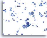
Figure 1
Bronchoalveolar lavage
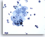
Low magnification showing an admixture of epithelial cells and macrophages. 20x
Bronchoalveolar lavage
Low magnification showing an admixture of epithelial cells and macrophages. 20x
Figure 1
Bronchoalveolar lavage
Low magnification showing an admixture of epithelial cells and macrophages.
20x
Bronchoalveolar lavage
Low magnification showing an admixture of epithelial cells and macrophages.
20x
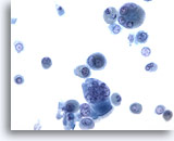
Figure 2
Bronchoalveolar lavage
Both macrophages and bronchial epithelial cells may be multinucleated. 40x
Bronchoalveolar lavage
Both macrophages and bronchial epithelial cells may be multinucleated. 40x
Figure 2
Bronchoalveolar lavage
Both macrophages and bronchial epithelial cells may be multinucleated.
40x
Bronchoalveolar lavage
Both macrophages and bronchial epithelial cells may be multinucleated.
40x
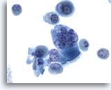
Figure 3
Bronchoalveolar lavage
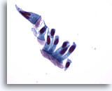
High magnification of a multinucleated bronchial epithelial cell. Note the terminal bar supporting cilia. 60x
Bronchoalveolar lavage
High magnification of a multinucleated bronchial epithelial cell. Note the terminal bar supporting cilia. 60x
Figure 3
Bronchoalveolar lavage
High magnification of a multinucleated bronchial epithelial cell. Note the terminal bar supporting cilia.
60x
Bronchoalveolar lavage
High magnification of a multinucleated bronchial epithelial cell. Note the terminal bar supporting cilia.
60x
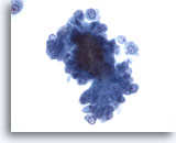
Figure 4
Bronchoalveolar lavage
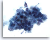
A cluster of benign bronchial epithelial cells may have a broad depth of focus. 40x
Bronchoalveolar lavage
A cluster of benign bronchial epithelial cells may have a broad depth of focus. 40x
Figure 4
Bronchoalveolar lavage
A cluster of benign bronchial epithelial cells may have a broad depth of focus.
40x
Bronchoalveolar lavage
A cluster of benign bronchial epithelial cells may have a broad depth of focus.
40x
Figure 5
Bronchoalveolar lavage
Oral squamous cells are common in respiratory specimens.
20x
Bronchoalveolar lavage
Oral squamous cells are common in respiratory specimens.
20x
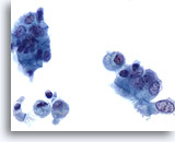
Figure 6
Bronchalveolar lavage
The columnar shape of bronchial epithelial cells, with basally oriented nuclei is illustrated in the cluster on the right. The size of these cells may vary significantly. 60x
Bronchalveolar lavage
The columnar shape of bronchial epithelial cells, with basally oriented nuclei is illustrated in the cluster on the right. The size of these cells may vary significantly. 60x
Figure 6
Bronchalveolar lavage
The columnar shape of bronchial epithelial cells, with basally oriented nuclei is illustrated in the cluster on the right. The size of these cells may vary significantly.
60x
Bronchalveolar lavage
The columnar shape of bronchial epithelial cells, with basally oriented nuclei is illustrated in the cluster on the right. The size of these cells may vary significantly.
60x
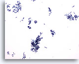
Figure 7
Bronchial brush
Low magnification illustrates many cell clusters collected by brushing. 10x
Bronchial brush
Low magnification illustrates many cell clusters collected by brushing. 10x
Figure 7
Bronchial brush
Low magnification illustrates many cell clusters collected by brushing.
10x
Bronchial brush
Low magnification illustrates many cell clusters collected by brushing.
10x
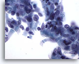
Figure 8
Bronchial brush
Although the cells are numerous, they maintain benign cytologic features 60x
Bronchial brush
Although the cells are numerous, they maintain benign cytologic features 60x
Figure 8
Bronchial brush
Although the cells are numerous, they maintain benign cytologic features
60x
Bronchial brush
Although the cells are numerous, they maintain benign cytologic features
60x
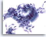
Figure 9
Bronchial brush
This cell cluster shows bronchial epithelial cells with cilia, a feature of benign bronchial epithelium. 40x
Bronchial brush
This cell cluster shows bronchial epithelial cells with cilia, a feature of benign bronchial epithelium. 40x
Figure 9
Bronchial brush
This cell cluster shows bronchial epithelial cells with cilia, a feature of benign bronchial epithelium.
40x
Bronchial brush
This cell cluster shows bronchial epithelial cells with cilia, a feature of benign bronchial epithelium.
40x
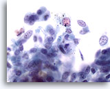
Figure 10
Bronchial brush
A single carbon-laden macrophage is shown adjacent to epithelial cells. 60x
Bronchial brush
A single carbon-laden macrophage is shown adjacent to epithelial cells. 60x
Figure 10
Bronchial brush
A single carbon-laden macrophage is shown adjacent to epithelial cells.
60x
Bronchial brush
A single carbon-laden macrophage is shown adjacent to epithelial cells.
60x
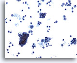
Figure 11
Bronchial wash
Low power magnification showing macrophages and a cluster of benign bronchial epithelial cells. 20x
Bronchial wash
Low power magnification showing macrophages and a cluster of benign bronchial epithelial cells. 20x
Figure 11
Bronchial wash
Low power magnification showing macrophages and a cluster of benign bronchial epithelial cells.
20x
Bronchial wash
Low power magnification showing macrophages and a cluster of benign bronchial epithelial cells.
20x
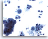
Figure 12
Bronchial wash
Compare the size of the cell cluster with adjacent macrophages and single epithelial cells. 40x
Bronchial wash
Compare the size of the cell cluster with adjacent macrophages and single epithelial cells. 40x
Figure 12
Bronchial wash
Compare the size of the cell cluster with adjacent macrophages and single epithelial cells.
40x
Bronchial wash
Compare the size of the cell cluster with adjacent macrophages and single epithelial cells.
40x
Figure 13
Bronchial wash
High magnification of the cluster shows cells with columnar shape.
60x
Bronchial wash
High magnification of the cluster shows cells with columnar shape.
60x
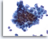
Figure 14
Bronchial wash
Although it may be difficult to see features of all cells, those at the periphery of the cluster have benign nuclear features 40x
Bronchial wash
Although it may be difficult to see features of all cells, those at the periphery of the cluster have benign nuclear features 40x
Figure 14
Bronchial wash
Although it may be difficult to see features of all cells, those at the periphery of the cluster have benign nuclear features
40x
Bronchial wash
Although it may be difficult to see features of all cells, those at the periphery of the cluster have benign nuclear features
40x
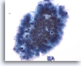
Figure 15
Bronchial wash
A cluster of bronchial epithelial cells may mimic adenocarcinoma. However, the cilia projecting from the cell periphery is a sign of their benign nature. 40x
Bronchial wash
A cluster of bronchial epithelial cells may mimic adenocarcinoma. However, the cilia projecting from the cell periphery is a sign of their benign nature. 40x
Figure 15
Bronchial wash
A cluster of bronchial epithelial cells may mimic adenocarcinoma. However, the cilia projecting from the cell periphery is a sign of their benign nature.
40x
Bronchial wash
A cluster of bronchial epithelial cells may mimic adenocarcinoma. However, the cilia projecting from the cell periphery is a sign of their benign nature.
40x
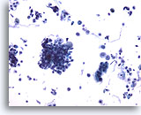
Figure 16
Bronchial wash
Low power magnification showing respiratory epithelial cells in a background of inflammation. 20x
Bronchial wash
Low power magnification showing respiratory epithelial cells in a background of inflammation. 20x
Figure 16
Bronchial wash
Low power magnification showing respiratory epithelial cells in a background of inflammation.
20x
Bronchial wash
Low power magnification showing respiratory epithelial cells in a background of inflammation.
20x
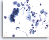
Figure 17
Bronchial wash
Polymorphonuclear leukocytes with macrophages and other material are present in the background. This may represent infection, chronic inflammatory conditions or other lung insults. 40x
Bronchial wash
Polymorphonuclear leukocytes with macrophages and other material are present in the background. This may represent infection, chronic inflammatory conditions or other lung insults. 40x
Figure 17
Bronchial wash
Polymorphonuclear leukocytes with macrophages and other material are present in the background. This may represent infection, chronic inflammatory conditions or other lung insults.
40x
Bronchial wash
Polymorphonuclear leukocytes with macrophages and other material are present in the background. This may represent infection, chronic inflammatory conditions or other lung insults.
40x
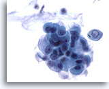
Figure 18
Bronchial wash
This cluster consists of bronchial epithelial cells and metaplastic cells. 60x
Bronchial wash
This cluster consists of bronchial epithelial cells and metaplastic cells. 60x
Figure 18
Bronchial wash
This cluster consists of bronchial epithelial cells and metaplastic cells.
60x
Bronchial wash
This cluster consists of bronchial epithelial cells and metaplastic cells.
60x
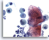
Figure 19
Bronchial wash
Bacteria from the oral cavity commonly contaminate the respiratory sample. Usually, they coat superficial oral squamous cells. 60x
Bronchial wash
Bacteria from the oral cavity commonly contaminate the respiratory sample. Usually, they coat superficial oral squamous cells. 60x
Figure 19
Bronchial wash
Bacteria from the oral cavity commonly contaminate the respiratory sample. Usually, they coat superficial oral squamous cells.
60x
Bronchial wash
Bacteria from the oral cavity commonly contaminate the respiratory sample. Usually, they coat superficial oral squamous cells.
60x
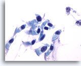
Figure 20
Sputum
Bronchial epithelial cells may be hyperchromatic or pyknotic in certain samples. 60X
Sputum
Bronchial epithelial cells may be hyperchromatic or pyknotic in certain samples. 60X
Figure 20
Sputum
Bronchial epithelial cells may be hyperchromatic or pyknotic in certain samples.
60X
Sputum
Bronchial epithelial cells may be hyperchromatic or pyknotic in certain samples.
60X
Figure 21
Sputum
High magnification showing bronchial epithelial cells with pyknotic nuclei.
60X
Sputum
High magnification showing bronchial epithelial cells with pyknotic nuclei.
60X

