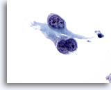
Fine Needle Aspiration Cytology – Pulmonary
PULMONARY
Rana S. Hoda MD, FIAC
Introduction
Lower respiratory cytology is important in the evaluation of pulmonary disease. Diagnosis is achieved by evaluation of exfoliative cytology (sputum, bronchial brushing, bronchial wash, and bronchoalveolar lavage) and fine needle aspiration (FNA) cytology (percutaneous, transthoracic, transbronchial and endoscopic-ultrasound guided).
For optimal pulmonary cytology proper specimen collection, fixation and processing are imperative. Collection in Cytolyt® for preparing a ThinPrep® slide has shown to be an excellent technique for respiratory tract specimens and has overcome many limitations of conventional smears by reducing the number of slides to examine, eliminating air-drying artifact and limiting obscuring elements such as mucus, blood and inflammation. Residual material from the specimen vial can be used for ancillary studies, such as special stains for organisms.
The main diagnostic issues in pulmonary cytology include identification of organisms, distinguishing reactive type 2 pneumocytes from well differentiated bronchioloalveolar carcinoma, reactive bronchial epithelium from malignancy, adenocarcinoma from squamous cell carcinoma, non-small cell carcinoma from small cell anaplastic carcinoma and cytologic differential diagnosis of neuroendocrine tumors. On ThinPrep slides microorganisms are easier to find and the morphology is similar to conventional smears. Due to well-preserved nuclear features the differential diagnosis between reactive type 2 pneumocytes and bronchioloalveolar carcinoma is relatively straightforward.
The general features of pulmonary cytology prepared by ThinPrep technique, compared to conventional preparation methods include:
Background
- Decreased inflammatory cells
- Decreased mucus
- Background material appears clumped or in aggregates rather than diffuse
Architectural Pattern:
- Uniform distribution of cells
- Smaller cell clusters, fragments and sheets
- Three-dimensional clusters can be seen
- Slightly more dispersed cell pattern
Cellular Features:
- Smaller cell size
- Well-preserved cytoplasmic features
- Well-preserved nuclei and nuclear detail
- Prominence of nucleoli
Nuclear molding in small cell anaplastic carcinoma is subtle. It appears more like gentle nuclear overlap. Crush artifact can be seen either as spindled nuclei or as long blue fibrous structures.
Advantages:
- Limited screening area
- Additional slides can be prepared for ancillary tests
- Reduction of obscuring elements
- Less overlap, even distribution, almost monolayer distribution
- Lack of air-drying
Bibliography
- Crapanzano JP, Zakowski MF: Diagnostic dilemmas in pulmonary cytology. Cancer (Cancer Cytopathol) 2001;93(6):364-375.
- Wiatrowska BA, Krol J, Zakowski MF: Large-cell neuroendocrine carcinoma of the lung: Proposed criteria for cytologic diagnosis. Diagn Cytopathol 2001; 24:58-64.
- Zimmerman RL, Montone KT, Fogt F, Norris AH: Ultra fast identification of Aspergillus species in pulmonary cytology specimens by in situ hybridization. Int J Mol Med 2000 Apr; 5(4): 427-9.
for an enlarged view.
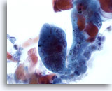
Pulmonary FNA, Herpes Simplex Virus Infection of Lung.
Characteristic viral cytopathic effects of multinucleation, nuclear molding, and ground-glass chromatin with eosinophilic intranuclear inclusions are seen. 60x
Pulmonary FNA, Herpes Simplex Virus Infection of Lung.
Characteristic viral cytopathic effects of multinucleation, nuclear molding, and ground-glass chromatin with eosinophilic intranuclear inclusions are seen.
60x
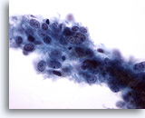
Pulmonary FNA, Large Cell Carcinoma of Lung. A sheet of poorly differentiated carcinoma cells with distinct cell borders. The cells are disorganized and show focal nuclear overlap. Background is clean and tumor diathesis is not seen. The tumor cells have no specific features of squamous or glandular differentiation. 40x
Pulmonary FNA, Large Cell Carcinoma of Lung.
A sheet of poorly differentiated carcinoma cells with distinct cell borders. The cells are disorganized and show focal nuclear overlap. Background is clean and tumor diathesis is not seen. The tumor cells have no specific features of squamous or glandular differentiation.
40x
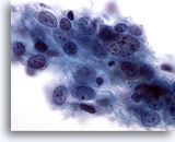
Pulmonary FNA, Large Cell Carcinoma of Lung.
The tumor cells are large when compared to the few apoptotic cells within the sheet. Cytoplasm is well-preserved, moderate in amount and dense to finely vacuolated. Cellular changes suggestive of intracellular bridges, a feature of squamous differentiation, are focally visible. Nuclei are round to oval with coarse clumped chromatin and thickened smooth membrane. Nucleoli are prominent, irregular, single and multiple. Nuclear to cytoplasmic ratio is high.
60x
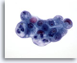
Pulmonary FNA, Adenocarcinoma of Lung. A cluster of malignant glandular cells with scalloped cell borders. Background is clean and tumor diathesis is not seen. The malignant cells are well-preserved. Cytoplasm shows fine to large discrete vacuoles, some with engulfed neutrophils. 60x
Pulmonary FNA, Adenocarcinoma of Lung.
A cluster of malignant glandular cells with scalloped cell borders. Background is clean and tumor diathesis is not seen. The malignant cells are well-preserved. Cytoplasm shows fine to large discrete vacuoles, some with engulfed neutrophils.
60x
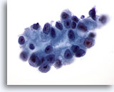
Pulmonary FNA, Adenocarcinoma of Lung.
Nuclei are round to oval, eccentric and have vesicular chromatin. Nuclear membrane shows some irregularity and thickness. Nucleoli are single, prominent and cherry-red in color. Nuclear to cytoplasmic ratio is high. 60x
Pulmonary FNA, Adenocarcinoma of Lung.
Nuclei are round to oval, eccentric and have vesicular chromatin. Nuclear membrane shows some irregularity and thickness. Nucleoli are single, prominent and cherry-red in color. Nuclear to cytoplasmic ratio is high.
60x
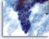
Pulmonary FNA, Bronchioloalveolar Carcinoma of Lung.
Three dimensional group of well differentiated adenocarcinoma cells with few ill-defined microacini. Background shows mucoid material. 60x
Pulmonary FNA, Bronchioloalveolar Carcinoma of Lung.
Three dimensional group of well differentiated adenocarcinoma cells with few ill-defined microacini. Background shows mucoid material.
60x
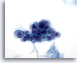
Pulmonary FNA, Bronchioloalveolar Carcinoma of Lung.
Cytoplasm is moderate in amount, of variable density with occasional vacuoles. Nuclei show loss of polarity, are small and hyperchromatic with minimal pleomorphism. These cells are difficult to distinguish from reactive bronchial cells. 60x
Pulmonary FNA, Bronchioloalveolar Carcinoma of Lung.
Cytoplasm is moderate in amount, of variable density with occasional vacuoles. Nuclei show loss of polarity, are small and hyperchromatic with minimal pleomorphism. These cells are difficult to distinguish from reactive bronchial cells.
60x
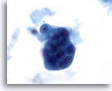
Pulmonary FNA, Bronchioloalveolar Carcinoma of Lung.
The three dimensional group shows considerable depth of focus, dense cytoplasm with hyperchromatic nuclei and prominent nucleoli. Background shows mucoid material. 60x
Pulmonary FNA, Bronchioloalveolar Carcinoma of Lung.
The three dimensional group shows considerable depth of focus, dense cytoplasm with hyperchromatic nuclei and prominent nucleoli. Background shows mucoid material.
60x
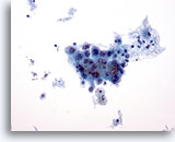
Pulmonary FNA, Poorly Differentiated Squamous Carcinoma of Lung.
A loosely cohesive fragment of malignant squamous cells is seen. Tumor diathesis appears as small clumps of necrotic material in the background. 20x
Pulmonary FNA, Poorly Differentiated Squamous Carcinoma of Lung.
A loosely cohesive fragment of malignant squamous cells is seen. Tumor diathesis appears as small clumps of necrotic material in the background.
20x
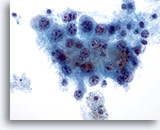
Pulmonary FNA, Poorly Differentiated Squamous Carcinoma of Lung. The cytoplasm is cyanophilic and densely vacuolated. Nuclei are enlarged, relatively round (compare the size with the nucleus of the benign macrophage in the lower mid field) with prominent nucleoli. Nuclear to cytoplasmic ratio is high. 40x
Pulmonary FNA, Poorly Differentiated Squamous Carcinoma of Lung.
The cytoplasm is cyanophilic and densely vacuolated. Nuclei are enlarged, relatively round (compare the size with the nucleus of the benign macrophage in the lower mid field) with prominent nucleoli. Nuclear to cytoplasmic ratio is high.
40x
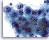
Pulmonary FNA, Poorly Differentiated Squamous Carcinoma of Lung. On higher magnification the cells show occasional intracellular bridges. Nuclei are centrally placed with coarse and unevenly distributed chromatin with prominent parachromatin clearing. Nucleoli are prominent and cherry-red in color. 60x
Pulmonary FNA, Poorly Differentiated Squamous Carcinoma of Lung.
On higher magnification the cells show occasional intracellular bridges. Nuclei are centrally placed with coarse and unevenly distributed chromatin with prominent parachromatin clearing. Nucleoli are prominent and cherry-red in color.
60x
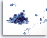
Pulmonary FNA, Poorly Differentiated Carcinoma of Lung. Scattered single cells and cohesive group of malignant cells with a high nuclear to cytoplasmic ratio. The cells around the periphery are better visualized. Cytoplasm is delicate and appears as a syncytium with sharp outer cell borders. 40x
Pulmonary FNA, Poorly Differentiated Carcinoma of Lung.
Scattered single cells and cohesive group of malignant cells with a high nuclear to cytoplasmic ratio. The cells around the periphery are better visualized. Cytoplasm is delicate and appears as a syncytium with sharp outer cell borders.
40x
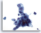
Pulmonary FNA, Poorly Differentiated Carcinoma of Lung.
Nuclei are round to oval, eccentric, hyperchromatic and show prominent nucleoli. The tumor cells lack definite cytological features of glandular or squamous origin 60x
Pulmonary FNA, Poorly Differentiated Carcinoma of Lung.
Nuclei are round to oval, eccentric, hyperchromatic and show prominent nucleoli. The tumor cells lack definite cytological features of glandular or squamous origin
60x
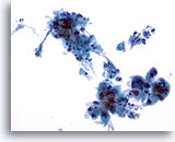
Pulmonary FNA, Non-Keratinizing Squamous Carcinoma of Lung.
Loosely cohesive sheet and single malignant squamous cells are seen in a background of clumps of necrosis. 20x
Pulmonary FNA, Non-Keratinizing Squamous Carcinoma of Lung.
Loosely cohesive sheet and single malignant squamous cells are seen in a background of clumps of necrosis.
20x
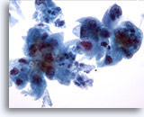
Pulmonary FNA, Non-Keratinizing Squamous Carcinoma of Lung.
Higher magnification shows a syncytium of cells with abundant dense cytoplasm with angulated and streaming borders. The nuclei are enlarged, hyperchromatic and show minimal overlap. 40x
Pulmonary FNA, Non-Keratinizing Squamous Carcinoma of Lung.
Higher magnification shows a syncytium of cells with abundant dense cytoplasm with angulated and streaming borders. The nuclei are enlarged, hyperchromatic and show minimal overlap.
40x
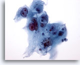
Pulmonary FNA, Non-Keratinizing Squamous Carcinoma of Lung. The malignant cells show dense cyanophilic cytoplasm with sharp cellular outlines. Few cells show intracytoplasmic vacuoles which may raise a diagnosis of adenocarcinoma. Nuclei have vesicular chromatin, minimal membrane irregularity and single or multiple prominent nucleoli. 60x
Pulmonary FNA, Non-Keratinizing Squamous Carcinoma of Lung.
The malignant cells show dense cyanophilic cytoplasm with sharp cellular outlines. Few cells show intracytoplasmic vacuoles which may raise a diagnosis of adenocarcinoma. Nuclei have vesicular chromatin, minimal membrane irregularity and single or multiple prominent nucleoli.
60x
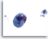
Pulmonary FNA, Non-Keratinizing Squamous Carcinoma of Lung.
Classic cell in cell arrangement of non-keratinizing squamous cell carcinoma is seen here. 60x
Pulmonary FNA, Non-Keratinizing Squamous Carcinoma of Lung.
Classic cell in cell arrangement of non-keratinizing squamous cell carcinoma is seen here.
60x
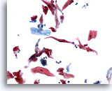
Pulmonary FNA, Keratinizing Squamous Carcinoma of Lung.
Dyshesive groups of keratinized squamous cells with orangeophilic, pleomorphic cytoplasm with elongated “fibre” and “tadpole” forms. 20x
Pulmonary FNA, Keratinizing Squamous Carcinoma of Lung.
Dyshesive groups of keratinized squamous cells with orangeophilic, pleomorphic cytoplasm with elongated “fibre” and “tadpole” forms.
20x

Pulmonary FNA, Keratinizing Squamous Carcinoma of Lung.
The backround shows a necrotic tumor diathesis. 20x
Pulmonary FNA, Keratinizing Squamous Carcinoma of Lung.
The backround shows a necrotic tumor diathesis.
20x
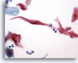
Pulmonary FNA, Keratinizing Squamous Carcinoma of Lung.
A single malignant cell with sharp straight cytoplasmic borders. 40x
Pulmonary FNA, Keratinizing Squamous Carcinoma of Lung.
A single malignant cell with sharp straight cytoplasmic borders.
40x
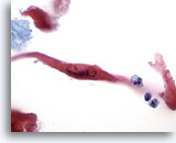
Pulmonary FNA, Keratinizing Squamous Carcinoma of Lung.
The nucleus is elongated, centrally placed with open chromatin, parachromatin clearing and a prominent nucleolus 60x
Pulmonary FNA, Keratinizing Squamous Carcinoma of Lung.
The nucleus is elongated, centrally placed with open chromatin, parachromatin clearing and a prominent nucleolus
60x
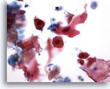
Pulmonary FNA, Keratinizing Squamous Carcinoma of Lung.
Single and small groups of keratinized squamous cells with background tumor diathesis. 40x
Pulmonary FNA, Keratinizing Squamous Carcinoma of Lung.
Single and small groups of keratinized squamous cells with background tumor diathesis.
40x
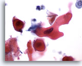
Pulmonary FNA, Keratinizing Squamous Carcinoma of Lung.
Higher maginification shows rigid pleomorphic cytoplasm and a single hyperchromatic nucleus with inconspicuous nucleoli. 60x
Pulmonary FNA, Keratinizing Squamous Carcinoma of Lung.
Higher maginification shows rigid pleomorphic cytoplasm and a single hyperchromatic nucleus with inconspicuous nucleoli.
60x
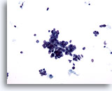
Pulmonary FNA, Small Cell Carcinoma of Lung.
Small clusters, many single hyperchromatic cells and few nuclei devoid of cytoplasm are seen. 20x
Pulmonary FNA, Small Cell Carcinoma of Lung.
Small clusters, many single hyperchromatic cells and few nuclei devoid of cytoplasm are seen.
20x
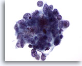
Pulmonary FNA, Small Cell Carcinoma of Lung.
Higher magnification shows a cluster of hyperchromatic cells with dual population. The viable cells show a high nuclear to cytoplasmic ratio with scant cytoplasm. Nuclei are hyperchromatic and irregular with “salt and pepper” chromatin. Nucleoli are not seen. Nuclear molding, although present, is subtle. The non-viable cells or individual tumor cell necrosis is seen as intermixed apoptotic bodies.
60x
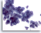
Pulmonary FNA, Small Cell Carcinoma of Lung. The scant cytoplasm appears as a thin rim around the nuclei. Chromatin appears granular and stippled in quality. Crush artifact is represented by a few spindled nuclei seen in the lower right hand corner instead of streaks of nuclear material. 60x
Pulmonary FNA, Small Cell Carcinoma of Lung.
The scant cytoplasm appears as a thin rim around the nuclei. Chromatin appears granular and stippled in quality. Crush artifact is represented by a few spindled nuclei seen in the lower right hand corner instead of streaks of nuclear material.
60x
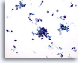
Pulmonary FNA, Small Cell Carcinoma of Lung.
Small clusters and many single hyperchromatic cells are seen in a background of tumor necrosis. 20x
Pulmonary FNA, Small Cell Carcinoma of Lung.
Small clusters and many single hyperchromatic cells are seen in a background of tumor necrosis.
20x
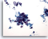
Pulmonary FNA, Small Cell Carcinoma of Lung.
Higher magnification shows cluster of hyperchromatic cells with dual population of viable cells and non-viable apoptotic cells. A few spindled cells towards the periphery (lower right hand corner) represent crush artifact. 40x
Pulmonary FNA, Small Cell Carcinoma of Lung.
Higher magnification shows cluster of hyperchromatic cells with dual population of viable cells and non-viable apoptotic cells. A few spindled cells towards the periphery (lower right hand corner) represent crush artifact.
40x
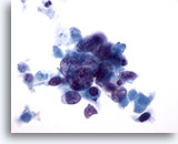
Pulmonary FNA, Small Cell Carcinoma of Lung.
Cytoplasm is scant and dense. Nuclei are round to oval and show molding. Nucleoli are not conspicuous. 60x
Pulmonary FNA, Small Cell Carcinoma of Lung.
Cytoplasm is scant and dense. Nuclei are round to oval and show molding. Nucleoli are not conspicuous.
60x
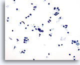
Pulmonary FNA, Small Cell Carcinoma of Lung.
Dyshesive groups and single hyperchromatic cells in a necrotic background 20x
Pulmonary FNA, Small Cell Carcinoma of Lung.
Dyshesive groups and single hyperchromatic cells in a necrotic background
20x
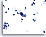
Pulmonary FNA, Small Cell Carcinoma of Lung.
Dual population of viable hyperchromatic cells and non-viable apoptotic cells are seen. Cytoplasm is scant and nuclear to cytoplasmic ratio is high. Nuclear molding is seen in the center of the field. 40x
Pulmonary FNA, Small Cell Carcinoma of Lung.
Dual population of viable hyperchromatic cells and non-viable apoptotic cells are seen. Cytoplasm is scant and nuclear to cytoplasmic ratio is high. Nuclear molding is seen in the center of the field.
40x
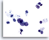
Pulmonary FNA, Small Cell Carcinoma of Lung.
Higher magnification shows the “salt and pepper chromatin” and absence of nucleoli. Some nuclei are devoid of cytoplasm. Focal spindled nuclear change (upper right hand corner) represents impending crush artifact. 60x
Pulmonary FNA, Small Cell Carcinoma of Lung.
Higher magnification shows the “salt and pepper chromatin” and absence of nucleoli. Some nuclei are devoid of cytoplasm. Focal spindled nuclear change (upper right hand corner) represents impending crush artifact.
60x
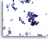
Pulmonary FNA, Intermediate Small Cell Carcinoma of Lung.
Small clusters and many single hyperchromatic cells are seen. Background shows tumor necrosis. 20x
Pulmonary FNA, Intermediate Small Cell Carcinoma of Lung.
Small clusters and many single hyperchromatic cells are seen. Background shows tumor necrosis.
20x
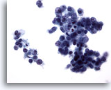
Pulmonary FNA, Intermediate Small Cell Carcinoma of Lung.
Higher magnification shows clusters of viable hyperchromatic cells with intermixed dark apoptotic bodies. Cytoplasm is scant and forms a thin rim around the nuclei. Nuclear cytoplasmic ratio is high. 40x
Pulmonary FNA, Intermediate Small Cell Carcinoma of Lung.
Higher magnification shows clusters of viable hyperchromatic cells with intermixed dark apoptotic bodies. Cytoplasm is scant and forms a thin rim around the nuclei. Nuclear cytoplasmic ratio is high.
40x
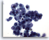
Pulmonary FNA, Intermediate Small Cell Carcinoma of Lung.
Viable nuclei show a single small nucleolus. This feature may make the distinction between intermediate small cell carcinoma and other poorly differentiated carcinomas and small cell squamous carcinoma difficult. The presence of a dual cell population, hyperchromatic cells with subtle nuclear molding and high nuclear and cytoplasmic ratio favors small cell carcinoma.
60x
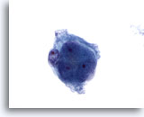
Pulmonary FNA, Metastatic Renal Cell Carcinoma of Lung.
Malignant renal carcinoma cells show clear cytoplasm, relatively bland nuclei with prominent nucleoli. 60x
Pulmonary FNA, Metastatic Renal Cell Carcinoma of Lung.
Malignant renal carcinoma cells show clear cytoplasm, relatively bland nuclei with prominent nucleoli.
60x
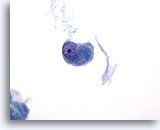
Pulmonary FNA, Metastatic Renal Cell Carcinoma of Lung.
Clinical history and multiple lung nodules with a “cannon ball” appearance favors a metatastic renal cell carcinoma. 60x
Pulmonary FNA, Metastatic Renal Cell Carcinoma of Lung.
Clinical history and multiple lung nodules with a “cannon ball” appearance favors a metatastic renal cell carcinoma.
60x
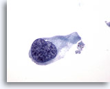
Chest wall FNA, Metastatic urothelial carcinoma.
The lesion yields single malignant cells with nuclei containing coarse hyperchromatic chromatin, similar to cells seen in urine specimens of patients with urothelial carcinoma. 60x
Chest wall FNA, Metastatic urothelial carcinoma.
The lesion yields single malignant cells with nuclei containing coarse hyperchromatic chromatin, similar to cells seen in urine specimens of patients with urothelial carcinoma.
60x
Chest wall FNA, Metastatic urothelial carcinoma.
Nucleoli are often present.
60x
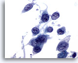
Chest wall FNA, Metastatic urothelial carcinoma.
The cytoplasm is dense and often shaped like a tadpole. 60x
Chest wall FNA, Metastatic urothelial carcinoma.
The cytoplasm is dense and often shaped like a tadpole.
60x

