Cytology of Pleural, Pericardial and Peritoneal Cavity Effusions - Metastatic Breast Cancer
EFFUSIONS – METASTATIC BREAST CANCER
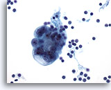
Advanced breast cancer metastatic to the liver and peritoneal cavity may induce a peritoneal effusion. The cancer cells are easily distinguished from inflammatory cells and benign mesothelial cells. As in pleural effusions, these cells tend to be present in large rounded clusters as well as individual cells.
Reminder: You may click on any slide image
for an enlarged view.
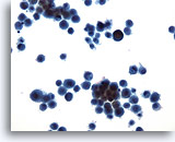
Peritoneal effusion: Metastatic carcinoma of the breast. Many small clusters and single malignant cells contrast with benign mesothelial cells. 20X
Peritoneal effusion:
Metastatic carcinoma of the breast. Many small clusters and single malignant cells contrast with benign mesothelial cells.
20X
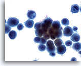
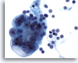
Peritoneal effusion: Metastatic carcinoma of the breast. Note cells with targetoid intracytoplasmic vacuoles containing mucin droplets. 40X
Metastatic carcinoma of the breast. Note cells with targetoid intracytoplasmic vacuoles containing mucin droplets.
40X
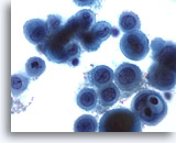
Peritoneal effusion: Metastatic carcinoma of the breast. Note malignant cells contrasting with benign histiocytes. Note large intracytoplasmic vacuoles with mucin droplets. 60X
Peritoneal effusion:
Metastatic carcinoma of the breast. Note malignant cells contrasting with benign histiocytes. Note large intracytoplasmic vacuoles with mucin droplets.
60X
Pleural effusion:
Metastatic ductal carcinoma
40X
Pleural effusion:
Metastatic ductal carcinoma
60X
Figures 73-74: Pleural effusion: Metastatic ductal carcinoma.
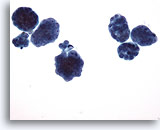
A cluster of metastatic ductal carcinoma cells exhibits large irregular nuclei and nucleoli with a background lymphocytes.
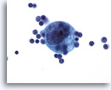
Pleural effusion: Metastatic ductal carcinoma. Note this single cell expressing nuclear gigantism. 60X
Pleural effusion:
Metastatic ductal carcinoma. Note this single cell expressing nuclear gigantism.
60X
Pleural effusion:
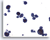
Metastatic carcinoma of the breast.
10X
Pleural effusion:
Metastatic carcinoma of the breast.
20X
Figures 76-77: Pleural effusion: Metastatic carcinoma of the breast.
The classic description of metastatic breast cancer in pleural effusions employs the term “cannonballs” to emphasize the rounded arrangement of tumor cells. Note the relatively small nuclear size. Nuclei are vesicular with prominent nucleoli. Cytoplasmic vacuoles are uncommon. A cell block of the cells allows for assay of hormonal receptors or other epithelial markers, such as her-2-neu.
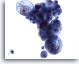
Peritoneal effusion: Metastatic carcinoma of the breast. Peritoneal effusion showing clusters of large tumor cells. The glandular nature of the tumor is obvious, but origin from breast is not easily determined. 40X
Peritoneal effusion:
Metastatic carcinoma of the breast. Peritoneal effusion showing clusters of large tumor cells. The glandular nature of the tumor is obvious, but origin from breast is not easily determined.
40X
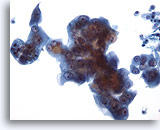
Pleural effusion: Metastatic carcinoma of the breast. Note two separate cell populations: a smaller benign population of mesothelial cells and larger tumor cells. 20X
Pleural effusion:
Metastatic carcinoma of the breast. Note two separate cell populations: a smaller benign population of mesothelial cells and larger tumor cells.
20X

