Respiratory Cytology - Dr. Jim Linder’s Bonus
DR. JIM LINDER’S BONUS:
OTHER RESPIRATORY STUFF
Reminder: You may click on any slide image
for an enlarged view.
for an enlarged view.

Figure 155
Ferruginous Body
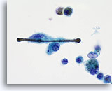
Fibers of asbestos, or other minerals may be coated with ferroprotein creating a “ferruginous body”. This structure is partially engulfed by a macrophage.
Ferruginous Body
Fibers of asbestos, or other minerals may be coated with ferroprotein creating a “ferruginous body”. This structure is partially engulfed by a macrophage.

Figure 156
Ferruginous Body
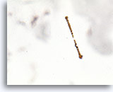
Ferruginous bodies due to asbestos fibers typically have a clear central core.
Ferruginous Body
Ferruginous bodies due to asbestos fibers typically have a clear central core.
Figure 156
Ferruginous Body
Ferruginous bodies due to asbestos fibers typically have a clear central core.
Ferruginous Body
Ferruginous bodies due to asbestos fibers typically have a clear central core.

Figure 157
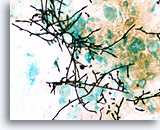
Candida spp.
Multiple hyphal forms of Candida spp. may be recovered in the respiratory samples of patients with oral candidiasis. (silver-based stain)
Candida spp.
Multiple hyphal forms of Candida spp. may be recovered in the respiratory samples of patients with oral candidiasis. (silver-based stain)

Figure 158
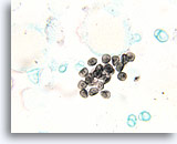
Pneumocystis carinii
The cyst forms of Pneumocystis carinii have a central dark zone when stained with silver-based stains.
Pneumocystis carinii
The cyst forms of Pneumocystis carinii have a central dark zone when stained with silver-based stains.
Figure 158
Pneumocystis carinii
The cyst forms of Pneumocystis carinii have a central dark zone when stained with silver-based stains.
Pneumocystis carinii
The cyst forms of Pneumocystis carinii have a central dark zone when stained with silver-based stains.

Figure 159
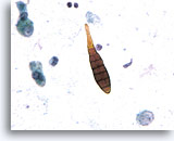
Alternaria spp
The macroconidia of Alternaria spp. has both longitudinal and septate hyphae.
Alternaria spp
The macroconidia of Alternaria spp. has both longitudinal and septate hyphae.
Figure 159
Alternaria spp.
The macroconidia of Alternaria spp. has both longitudinal and septate hyphae.
Alternaria spp.
The macroconidia of Alternaria spp. has both longitudinal and septate hyphae.

Figure 160
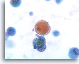
Pollen
Pollen granules have a thick refractile wall, multiple surface spikes, and are typically pigmented.
Pollen
Pollen granules have a thick refractile wall, multiple surface spikes, and are typically pigmented.
Figure 160
Pollen
Pollen granules have a thick refractile wall, multiple surface spikes, and are typically pigmented.
Pollen
Pollen granules have a thick refractile wall, multiple surface spikes, and are typically pigmented.

Figure 161
Macrophages
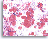
In cases of lipoid, or aspiration pneumonia the macrophages contain multiple complex lipid vacuoles.
Macrophages
In cases of lipoid, or aspiration pneumonia the macrophages contain multiple complex lipid vacuoles.
Figure 161
Macrophages
In cases of lipoid, or aspiration pneumonia the macrophages contain multiple complex lipid vacuoles.
Macrophages
In cases of lipoid, or aspiration pneumonia the macrophages contain multiple complex lipid vacuoles.
Figure 162
Lipid
The lipid in cases of lipoid pneumonia may be highlighted by oil-red O stain.
Lipid
The lipid in cases of lipoid pneumonia may be highlighted by oil-red O stain.

Figure 163
Macrophages
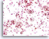
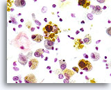
Patients who sustain injury to the microvasculature of the lung develop pulmonary hemorrhage, manifested by hemosiderin-containing macrophages.
Macrophages
Patients who sustain injury to the microvasculature of the lung develop pulmonary hemorrhage, manifested by hemosiderin-containing macrophages.
Figure 164
Lymphocytosis
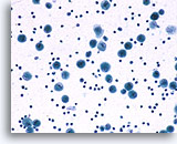
Patients with sarcoidosis typically have an interstitial lymphocytosis.
Lymphocytosis
Patients with sarcoidosis typically have an interstitial lymphocytosis.

Figure 165
Epithelial Cells
Highly reactive epithelial cells may be seen in patients with pulmonary aspergilloma.
Epithelial Cells
Highly reactive epithelial cells may be seen in patients with pulmonary aspergilloma.
Figure 165
Epithelial Cells
Highly reactive epithelial cells may be seen in patients with pulmonary aspergilloma.
Epithelial Cells
Highly reactive epithelial cells may be seen in patients with pulmonary aspergilloma.

Figure 166
Cryptococcus
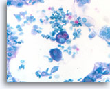
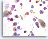
Multiple intracellular yeasts are seen within the cytoplasm of macrophages in Cryptococcus infection.
Cryptococcus
Multiple intracellular yeasts are seen within the cytoplasm of macrophages in Cryptococcus infection.
Figure 166
Cryptococcus
Multiple intracellular yeasts are seen within the cytoplasm of macrophages in Cryptococcus infection.
Cryptococcus
Multiple intracellular yeasts are seen within the cytoplasm of macrophages in Cryptococcus infection.

Figure 167
Cryptococcus neoformans
The capsule of Cryptococcus neoformans may be seen with mucin based stains.
Cryptococcus neoformans
The capsule of Cryptococcus neoformans may be seen with mucin based stains.
Figure 167
Cryptococcus neoformans
The capsule of Cryptococcus neoformans may be seen with mucin based stains.
Cryptococcus neoformans
The capsule of Cryptococcus neoformans may be seen with mucin based stains.

Figure 168
Ciliocytophthoria
Respiratory viral infections may cause tufts of cilia to become detached from bronchial epithelial cells. This process is termed ciliocytophthoria.
Ciliocytophthoria
Respiratory viral infections may cause tufts of cilia to become detached from bronchial epithelial cells. This process is termed ciliocytophthoria.

Figure 169
Hyperplasia
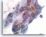
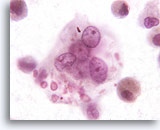
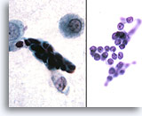
Reserve cell hyperplasia is characterized by tight clusters of small reserve cells.
Hyperplasia
Reserve cell hyperplasia is characterized by tight clusters of small reserve cells.
Figure 169
Hyperplasia
Reserve cell hyperplasia is characterized by tight clusters of small reserve cells.
Hyperplasia
Reserve cell hyperplasia is characterized by tight clusters of small reserve cells.

