Respiratory Cytology - Adenocarcinoma
ADENOCARCINOMA
Reminder: You may click on any slide image
for an enlarged view.
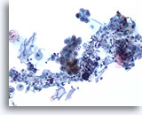
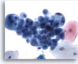
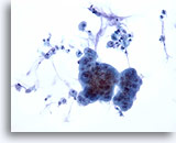
Figure 105
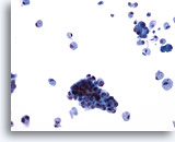
Bronchial wash
The cells of well and moderately-differentiated pulmonary adenocarcinoma are typically clustered. 20x
Bronchial wash
The cells of well and moderately-differentiated pulmonary adenocarcinoma are typically clustered. 20x
Figure 105
Bronchial wash
The cells of well and moderately-differentiated pulmonary adenocarcinoma are typically clustered.
20x
Bronchial wash
The cells of well and moderately-differentiated pulmonary adenocarcinoma are typically clustered.
20x
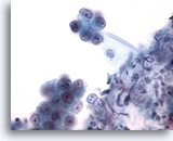
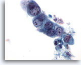
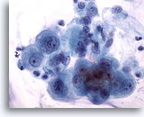
Figure 106
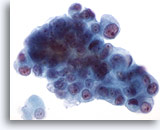
Bronchial wash
Higher magnification shows relatively bland uniform nuclei in the cell cluster. 40x
Bronchial wash
Higher magnification shows relatively bland uniform nuclei in the cell cluster. 40x
Figure 106
Bronchial wash
Higher magnification shows relatively bland uniform nuclei in the cell cluster.
40x
Bronchial wash
Higher magnification shows relatively bland uniform nuclei in the cell cluster.
40x
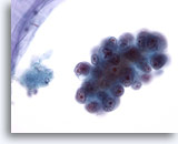
Figure 107
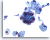
Bronchial wash
A cellular feature of adenocarcinoma is that the nucleoli are usually single and centrally placed. 40x
Bronchial wash
A cellular feature of adenocarcinoma is that the nucleoli are usually single and centrally placed. 40x
Figure 107
Bronchial wash
A cellular feature of adenocarcinoma is that the nucleoli are usually single and centrally placed.
40x
Bronchial wash
A cellular feature of adenocarcinoma is that the nucleoli are usually single and centrally placed.
40x
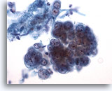
Figure 108
Bronchial wash
The three-dimensional nature of this cluster of adenocarcinoma is apparent. 40x
Bronchial wash
The three-dimensional nature of this cluster of adenocarcinoma is apparent. 40x
Figure 108
Bronchial wash
The three-dimensional nature of this cluster of adenocarcinoma is apparent.
40x
Bronchial wash
The three-dimensional nature of this cluster of adenocarcinoma is apparent.
40x
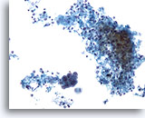
Figure 109
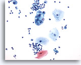
Bronchial wash
A cluster of adenocarcinoma is admixed with respiratory macrophages.
20x
Bronchial wash
A cluster of adenocarcinoma is admixed with respiratory macrophages.
20x
Figure 110
Bronchial wash
Cells with vacuolated cytoplasm are seen at the edge of the cluster.
60x
Bronchial wash
Cells with vacuolated cytoplasm are seen at the edge of the cluster.
60x
Figure 111
Bronchial wash
This group of cells forms a small acinar structure. 60x
Bronchial wash
This group of cells forms a small acinar structure. 60x
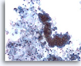
Figure 112
Bronchial wash
This cluster of adenocarcinoma is present in a background of necrosis. 20x
Bronchial wash
This cluster of adenocarcinoma is present in a background of necrosis. 20x
Figure 112
Bronchial wash
This cluster of adenocarcinoma is present in a background of necrosis.
20x
Bronchial wash
This cluster of adenocarcinoma is present in a background of necrosis.
20x
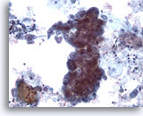
Figure 113
Bronchial wash
When much of the material is necrotic, the diagnosis of adenocarcinoma should be made with great caution. 20x
Bronchial wash
When much of the material is necrotic, the diagnosis of adenocarcinoma should be made with great caution. 20x
Figure 113
Bronchial wash
When much of the material is necrotic, the diagnosis of adenocarcinoma should be made with great caution.
20x
Bronchial wash
When much of the material is necrotic, the diagnosis of adenocarcinoma should be made with great caution.
20x
Figure 114
Sputum
This sputum sample shows a cluster of vacuolated cells.
20x
Sputum
This sputum sample shows a cluster of vacuolated cells.
20x
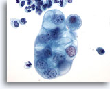
Figure 115
Sputum
Higher magnification shows the multiple complex vacuoles within the cytoplasm. 60x
Sputum
Higher magnification shows the multiple complex vacuoles within the cytoplasm. 60x
Figure 115
Sputum
Higher magnification shows the multiple complex vacuoles within the cytoplasm.
60x
Sputum
Higher magnification shows the multiple complex vacuoles within the cytoplasm.
60x
Figure 116
Sputum
A well-formed acinar structure is present within this cluster
60x
Sputum
A well-formed acinar structure is present within this cluster
60x
Figure 117
Bronchial wash
A more poorly-differentiated adenocarcinoma contains cell debris.
20x
Bronchial wash
A more poorly-differentiated adenocarcinoma contains cell debris.
20x
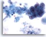
Figure 118
Bronchial wash
High magnification of the preserved cells shows features of adenocarcinoma: cell clustering, cell monotony, pale chromatin and eosinophilic nucleoli. 60x
Bronchial wash
High magnification of the preserved cells shows features of adenocarcinoma: cell clustering, cell monotony, pale chromatin and eosinophilic nucleoli. 60x
Figure 118
Bronchial wash
High magnification of the preserved cells shows features of adenocarcinoma: cell clustering, cell monotony, pale chromatin and eosinophilic nucleoli.
60x
Bronchial wash
High magnification of the preserved cells shows features of adenocarcinoma: cell clustering, cell monotony, pale chromatin and eosinophilic nucleoli.
60x
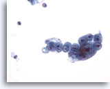
Figure 119
Bronchial wash
Note the centrally located nucleoli and the rudimentary acinar structure in this adenocarcinoma. 40x
Bronchial wash
Note the centrally located nucleoli and the rudimentary acinar structure in this adenocarcinoma. 40x
Figure 119
Bronchial wash
Note the centrally located nucleoli and the rudimentary acinar structure in this adenocarcinoma.
40x
Bronchial wash
Note the centrally located nucleoli and the rudimentary acinar structure in this adenocarcinoma.
40x
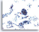
Figure 120
Bronchoalveolar lavage
Low power magnification shows a poorly-differentiated adenocarcinoma. 20x
Bronchoalveolar lavage
Low power magnification shows a poorly-differentiated adenocarcinoma. 20x
Figure 120
Bronchoalveolar lavage
Low power magnification shows a poorly-differentiated adenocarcinoma.
20x
Bronchoalveolar lavage
Low power magnification shows a poorly-differentiated adenocarcinoma.
20x
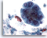
Figure 121
Bronchoalveolar lavage
The nuclei from this poorly-differentiated adenocarcinoma are variable in shape and have coarsely clumped chromatin. 60x
Bronchoalveolar lavage
The nuclei from this poorly-differentiated adenocarcinoma are variable in shape and have coarsely clumped chromatin. 60x
Figure 121
Bronchoalveolar lavage
The nuclei from this poorly-differentiated adenocarcinoma are variable in shape and have coarsely clumped chromatin.
60x
Bronchoalveolar lavage
The nuclei from this poorly-differentiated adenocarcinoma are variable in shape and have coarsely clumped chromatin.
60x
Figure 122
Bronchoalveolar lavage
Poorly-differentiated adenocarcinoma.
60x
Bronchoalveolar lavage
Poorly-differentiated adenocarcinoma.
60x
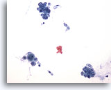
Figure 123
Bronchial wash
Numerous cell clusters are present in this adenocarcinoma of terminal bronchiolar origin.
20x
Bronchial wash
Numerous cell clusters are present in this adenocarcinoma of terminal bronchiolar origin.
20x
Figure 123
Bronchial wash
Numerous cell clusters are present in this adenocarcinoma of terminal bronchiolar origin.
20x
Bronchial wash
Numerous cell clusters are present in this adenocarcinoma of terminal bronchiolar origin.
20x
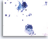
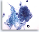
Figure 124
Bronchial wash
Compare the size of the cells in the cluster to the adjacent columnar epithelium. 20x
Bronchial wash
Compare the size of the cells in the cluster to the adjacent columnar epithelium. 20x
Figure 124
Bronchial wash
Compare the size of the cells in the cluster to the adjacent columnar epithelium.
20x
Bronchial wash
Compare the size of the cells in the cluster to the adjacent columnar epithelium.
20x
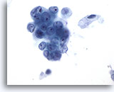
Figure 125
Bronchial wash
High magnification showing adenocarcinoma of terminal bronchiolar origin. 60x
Bronchial wash
High magnification showing adenocarcinoma of terminal bronchiolar origin. 60x
Figure 125
Bronchial wash
High magnification showing adenocarcinoma of terminal bronchiolar origin.
60x
Bronchial wash
High magnification showing adenocarcinoma of terminal bronchiolar origin.
60x
Figure 126
Bronchial wash
Adenocarcinoma
60x
Bronchial wash
Adenocarcinoma
60x
Figure 127
Bronchial wash
Bronchial wash with malignant cells of terminal bronchiolar origin.
20x
Bronchial wash
Bronchial wash with malignant cells of terminal bronchiolar origin.
20x
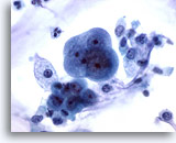
Figure 128
Bronchial wash
High magnification shows the relatively bland features in cells of this adenocarcinoma of terminal bronchiolar origin. 60x
Bronchial wash
High magnification shows the relatively bland features in cells of this adenocarcinoma of terminal bronchiolar origin. 60x
Figure 128
Bronchial wash
High magnification shows the relatively bland features in cells of this adenocarcinoma of terminal bronchiolar origin.
60x
Bronchial wash
High magnification shows the relatively bland features in cells of this adenocarcinoma of terminal bronchiolar origin.
60x
Figure 129
Bronchial wash
Adenocarcinoma of terminal bronchiolar origin. 60x
Bronchial wash
Adenocarcinoma of terminal bronchiolar origin. 60x
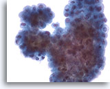
Figure 130
Bronchial brushing
Adenocarcinoma may show true papillary or pseudo-papillary structures as seen in this case. 40x
Bronchial brushing
Adenocarcinoma may show true papillary or pseudo-papillary structures as seen in this case. 40x
Figure 130
Bronchial brushing
Adenocarcinoma may show true papillary or pseudo-papillary structures as seen in this case.
40x
Bronchial brushing
Adenocarcinoma may show true papillary or pseudo-papillary structures as seen in this case.
40x

