Reminder: You may click on any slide image
for an enlarged view.
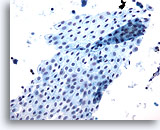
Figure 13
Peritoneal wash: Benign. 20X
Figure 13
Peritoneal wash:
Benign.
20X
For peritoneal washings the surgeon performs an irrigation or barbitage to collect cells for analysis. Pelvic mesothelial cells may be shed in broad and flat sheets. Sometimes the sheets fold so that overlapping of cells is evident. The benign nature of these mesothelial cells is obvious by their uniform arrangement and appearance.
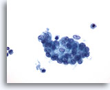
Figure 14
Peritoneal wash: Benign mesothelial cells in a cluster. 40X
Figure 14
Peritoneal wash:
Benign mesothelial cells in a cluster.
40X
Benign mesothelial cells from a peritoneal washing may also appear rounded up into a more three-dimensional group with some overlap. Close examination reveals uniform cells with benign nuclear features.
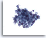
Figure 15
Peritoneal wash: Reactive mesothelial cells. 40X
Figure 15
Peritoneal wash:
Reactive mesothelial cells.
40X
This group of benign mesothelial cells also demonstrates slight overlapping. Clusters of benign mesothelial cells have a scalloped, or hobnail border, in contrast to the smooth border typically found in clusters of adenocarcinoma.
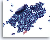
Figure 16
Pelvic wash: Benign mesothelial cells. 20X
Figure 16
Pelvic wash:
Benign mesothelial cells.
20X
A flat sheet of evenly spaced mesothelial cells in a honeycombed pattern.
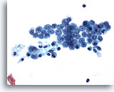
Figure 17
Pelvic wash: Benign mesothelial cells. 40X
Figure 17
Pelvic wash:
Benign mesothelial cells.
40X
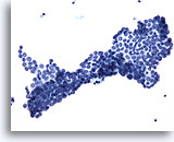
Figure 18
Pelvic wash: Benign mesothelial cells. 20X
Figure 18
Pelvic wash:
Benign mesothelial cells.
20X
Figures 17 and 18 show large sheets of mesothelial cells. Nuclei are vesicular and uniform with small or inconspicuous nucleoli.
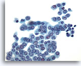
Figure 19
Pelvic wash: Negative. 40X
Figure 19
Pelvic wash:
Negative.
40X
Mesothelial cells can also have a cuboidal appearance with more condensed cytoplasm. Again, note the cohesive sheet of cells with uniformity of nuclei throughout.
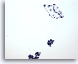
Figure 20
Peritoneal wash: Artifacts. 20X
Figure 20
Peritoneal wash:
Artifacts.
20X
Cholesterol and other types of crystals may be seen in peritoneal washes and fluids from other body sites.
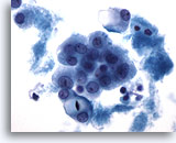
Figure 21
Peritoneal wash: Artifacts. 60X
Figure 21
Peritoneal wash:
Artifacts.
60X
Reactive mesothelial cells containing starch granules are indicative of glove powder contamination from a prior laparotomy. Starch granules may also contaminate the specimen via the gloves of the technician processing the specimen.