Reminder: You may click on any slide image
for an enlarged view.
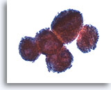
Figure 22
Pleural effusion Mesothelioma. 10X
Figure 22
Pleural effusion:
Mesothelioma.
10X
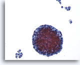
Figure 23
Pleural effusion Mesothelioma. 20X
Figure 23
Pleural effusion:
Mesothelioma.
20X
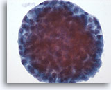
Figure 24
Pleural effusion Mesothelioma. 40X
Figure 24
Pleural effusion:
Mesothelioma.
40X
Figures 22-24:
Various magnifications show “cannon-ball” masses of tumor cells. The key to diagnosing mesothelioma is not identifying a second malignant cell population. Final determination may require immunocytochemistry or a cell block with immunohistochemistry, electron microscopy, or other specialized techniques.
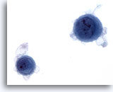
Figure 25
Pleural effusion Mesothelioma. 60X
Figure 25
Pleural effusion:
Mesothelioma.
60X
Individual malignant mesothelial cells exhibit a rim of ruffled, less dense cytoplasm (ectoplasm), surrounding dense cytoplasm around the nucleus (endoplasm).
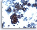
Figure 26
Pleural effusion Mesothelioma. 20X
Figure 26
Pleural effusion:
Mesothelioma.
20X
Tumor cells may be seen in a background of blood and proteinaceous debris. Groups of more than 12 cells may be a feature of malignancy.
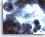
Figure 27
Pleural effusion Mesothelioma. 60X
Figure 27
Pleural effusion:
Mesothelioma.
60X
At higher magnification, the high N/C ratio with variability in nuclear size and occasional multi-nucleation confirm the malignant nature of these cells. Differential diagnoses include adenocarcinoma and mesothelioma. Fine microscopic features of peripheral cell membranes and intercellular windows may suggest mesothelioma.
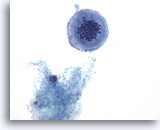
Figure 28
Pleural effusion Mesothelioma. 60X
Figure 28
Pleural effusion:
Mesothelioma.
60X
Abnormal mitotic figures may be noted with mesothelioma, other malignancies, as well as occasional reactive mesothelial cells in effusions.