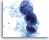
Cytology of Pleural, Pericardial and Peritoneal Cavity Effusions - Effusions and Washes Other Gynecologic Metastases
EFFUSIONS AND WASHES – OTHER GYNECOLOGIC METASTASES
Reminder: You may click on any slide image
for an enlarged view.
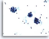
Peritoneal effusion:
Papillary serous carcinoma of the endometrium.
20X
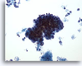
Peritoneal effusion:
Papillary serous carcinoma of the endometrium.
40X
Figures 55-56:
Peritoneal effusion, papillary serous carcinoma of the endometrium. Malignant cells clustered around psammoma bodies.
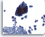
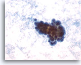
Peritoneal effusion:
Endometrial adenocarcinoma. Low power photomicrograph shows two separate cell populations. Note the flat sheet at the lower left of uniform, benign cells, many dyshesive tumor cells at lower right, with a large cohesive fragment of tumor cells at upper left.
20X
Peritoneal effusion:
Endometrial adenocarcinoma. Low power photomicrograph shows two separate cell populations. Note the flat sheet at the lower left of uniform, benign cells, many dyshesive tumor cells at lower right, with a large cohesive fragment of tumor cells at upper left.
20X
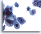
Peritoneal effusion: Endometrial adenocarcinoma. Malignant cells with features of adenocarcinoma including a mitotic figure, prominent nucleoli, vacuolated cytoplasm and eccentric nuclei. 60X
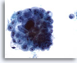
Peritoneal effusion:
Endometrial adenocarcinoma. Malignant cells with features of adenocarcinoma including a mitotic figure, prominent nucleoli, vacuolated cytoplasm and eccentric nuclei.
60X
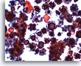
Pleural effusion:
Endometrial adenocarcinoma.
20X
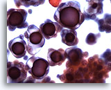
Pleural effusion:
Endometrial adenocarcinoma.
40X
Figures 59-60:
Pleural effusion: Endometrial adenocarcinoma.
This pleural effusion contains large clusters of malignant cells. Note increased cell size relative to lymphocytes and mesothelial cells in the background. A discontinuous population is one characteristic of malignancy in body effusions
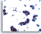
Pleural effusion: Papillary adenocarcinoma of peritoneal surface. The origin of this tumor is extra-ovarian carcinoma or carcinoma of Müellerian origin. Note distinction from benign cells. 10X
Pleural effusion:
Papillary adenocarcinoma of peritoneal surface. The origin of this tumor is extra-ovarian carcinoma or carcinoma of Müellerian origin. Note distinction from benign cells.
10X
Pleural effusion:
Adenocarcinoma. Papillary clusters of malignant cells.
20X
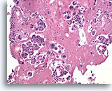
Pleural effusion cell block: Adenocarcinoma. A cell block further highlights the distinct malignant cell population and allows for immunohistochemical analysis of cell markers for differentiation and prognosis. 10X
Pleural effusion cell block:
Adenocarcinoma. A cell block further highlights the distinct malignant cell population and allows for immunohistochemical analysis of cell markers for differentiation and prognosis.
10X
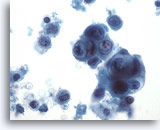
Pelvic wash: Undifferentiated large cell carcinoma of the cervix. Cytology shows rounded clusters of malignant cells. 60X
Pelvic wash:
Undifferentiated large cell carcinoma of the cervix. Cytology shows rounded clusters of malignant cells.
60X
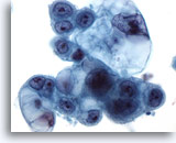
Pelvic wash: Carcinoma of the cervix. Magnified view shows vacuolated cytoplasm, prominent nucleoli, and powdery chromatin pattern suggesting an adenocarcinoma. 60X
Pelvic wash:
Carcinoma of the cervix. Magnified view shows vacuolated cytoplasm, prominent nucleoli, and powdery chromatin pattern suggesting an adenocarcinoma.
60X
Pelvic wash:
Carcinosarcoma.
20X
Pelvic wash:
Carcinosarcoma.
60X
Figures 66-67:
Pelvic wash: Carcinosarcoma.
Clusters of high-grade malignant cells. Look for both malignant epithelial and malignant stromal cell components.
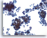
Peritoneal effusion:
Fallopian tube adenocarcinoma.
20X
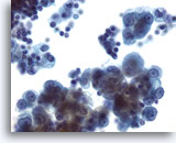
Peritoneal effusion:
Fallopian tube adenocarcinoma.
60X
Figures 68-69:
Peritoneal effusion: Fallopian tube adenocarcinoma.
A rare, highly malignant tumor. Consider this diagnosis when peritoneal effusion presents without evidence of ovarian or endometrial pathology. Note the prominent cytoplasmic vacuoles.

