Urinary Tract Cytology - Low Grade Urothelial Carcinoma
LOW GRADE UROTHELIAL CARCINOMA AND CARCINOMA IN SITU
Reminder: You may click on any slide image
for an enlarged view.
for an enlarged view.

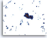
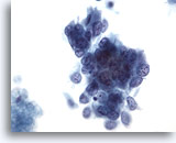
Figure 43
Voided urine, low grade carcinoma
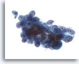
Low grade urothelial carcinoma is characterized by increased clusters of cells. 20x
Figure 43
Voided urine, low grade carcinoma
Low grade urothelial carcinoma is characterized by increased clusters of cells.
20x
Voided urine, low grade carcinoma
Low grade urothelial carcinoma is characterized by increased clusters of cells.
20x

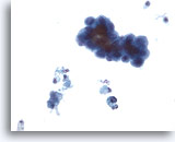
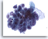
Figure 44
Voided urine, low grade carcinoma
The clusters in low grade urothelial carcinoma may or may not be papillary. 40x
Figure 44
Voided urine, low grade carcinoma
The clusters in low grade urothelial carcinoma may or may not be papillary.
40x
Voided urine, low grade carcinoma
The clusters in low grade urothelial carcinoma may or may not be papillary.
40x

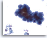
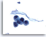
Figure 45
Voided urine, low grade carcinoma
The cells in the clusters of low grade urothelial carcinoma have high N/C ratios. 60x
Voided urine, low grade carcinoma
The cells in the clusters of low grade urothelial carcinoma have high N/C ratios. 60x
Figure 45
Voided urine, low grade carcinoma
The cells in the clusters of low grade urothelial carcinoma have high N/C ratios.
60x
Voided urine, low grade carcinoma
The cells in the clusters of low grade urothelial carcinoma have high N/C ratios.
60x

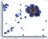
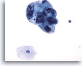
Figure 46
Voided urine, low grade carcinoma
Nuclei in low grade urothelial carcinoma sometimes bulge out of the cytoplasm. 40x
Figure 46
Voided urine, low grade carcinoma
Nuclei in low grade urothelial carcinoma sometimes bulge out of the cytoplasm.
40x
Voided urine, low grade carcinoma
Nuclei in low grade urothelial carcinoma sometimes bulge out of the cytoplasm.
40x

Figure 47
Bladder urine, low grade carcinoma
A loose cluster of cells from low grade urothelial carcinoma can be compared to reactive urothelial cells. 20x
Bladder urine, low grade carcinoma
A loose cluster of cells from low grade urothelial carcinoma can be compared to reactive urothelial cells. 20x

Figure 48
Bladder urine, low grade carcinoma
Nuclei of low grade urothelial carcinoma are irregular and may appear to have notches or grooves. 40x
Bladder urine, low grade carcinoma
Nuclei of low grade urothelial carcinoma are irregular and may appear to have notches or grooves. 40x
Figure 48
Bladder urine, low grade carcinoma
Nuclei of low grade urothelial carcinoma are irregular and may appear to have notches or grooves.
40x
Bladder urine, low grade carcinoma
Nuclei of low grade urothelial carcinoma are irregular and may appear to have notches or grooves.
40x

Figure 49
Cystoscopy urine, low grade carcinoma
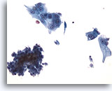
Compare the normal urothelial on the right to the crowded cluster on the left. 40x
Cystoscopy urine, low grade carcinoma
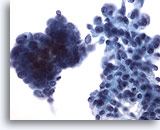
Compare the normal urothelial on the right to the crowded cluster on the left. 40x
Figure 49
Cystoscopy urine, low grade carcinoma
Compare the normal urothelial on the right to the crowded cluster on the left.
40x
Cystoscopy urine, low grade carcinoma
Compare the normal urothelial on the right to the crowded cluster on the left.
40x

Figure 50
Urine, low grade carcinoma
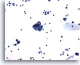
Cells of low grade urothelial carcinoma may be in small groups with some single cells. 20x
Urine, low grade carcinoma
Cells of low grade urothelial carcinoma may be in small groups with some single cells. 20x
Figure 50
Urine, low grade carcinoma
Cells of low grade urothelial carcinoma may be in small groups with some single cells.
20x
Urine, low grade carcinoma
Cells of low grade urothelial carcinoma may be in small groups with some single cells.
20x

Figure 51
Urine, low grade carcinoma
Nucleoli are usually indistinct or absent in low grade urothelial carcinoma. 40x
Urine, low grade carcinoma
Nucleoli are usually indistinct or absent in low grade urothelial carcinoma. 40x
Figure 51
Urine, low grade carcinoma
Nucleoli are usually indistinct or absent in low grade urothelial carcinoma.
40x
Urine, low grade carcinoma
Nucleoli are usually indistinct or absent in low grade urothelial carcinoma.
40x

Figure 52
Voided urine, low grade carcinoma
Chromatin in low grade urothelial carcinoma is usually granular and evenly distributed. 60x
Voided urine, low grade carcinoma
Chromatin in low grade urothelial carcinoma is usually granular and evenly distributed. 60x
Figure 52
Voided urine, low grade carcinomaChromatin in low grade urothelial carcinoma is usually granular and evenly distributed.
60x
Voided urine, low grade carcinomaChromatin in low grade urothelial carcinoma is usually granular and evenly distributed.
60x

Figure 53
Urine, urothelial carcinoma in situ
Cytologic features of carcinoma in situ are similar to those of high grade urothelial carcinoma. 60x
Urine, urothelial carcinoma in situ
Cytologic features of carcinoma in situ are similar to those of high grade urothelial carcinoma. 60x
Figure 53
Urine, urothelial carcinoma in situ
Cytologic features of carcinoma in situ are similar to those of high grade urothelial carcinoma.
60x
Urine, urothelial carcinoma in situ
Cytologic features of carcinoma in situ are similar to those of high grade urothelial carcinoma.
60x

Figure 54
Urine, urothelial carcinoma in situ
Invasion can be suspected when tumor cells are associated with diathesis, but an unequivocal diagnosis of CIS versus invasion cannot be rendered by cytology alone. 40x
Urine, urothelial carcinoma in situ
Invasion can be suspected when tumor cells are associated with diathesis, but an unequivocal diagnosis of CIS versus invasion cannot be rendered by cytology alone. 40x
Figure 54
Urine, urothelial carcinoma in situ
Invasion can be suspected when tumor cells are associated with diathesis, but an unequivocal diagnosis of CIS versus invasion cannot be rendered by cytology alone.
40x
Urine, urothelial carcinoma in situ
Invasion can be suspected when tumor cells are associated with diathesis, but an unequivocal diagnosis of CIS versus invasion cannot be rendered by cytology alone.
40x
