Respiratory Cytology - Squamous Cell Carcinoma
SQUAMOUS CELL CARCINOMA
Reminder: You may click on any slide image
for an enlarged view.
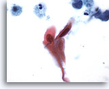
Figure 76
Sputum
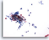
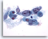
Orangeophilic cells with hyperchromatic nuclei are suspicious for squamous cell carcinoma. 60x
Sputum
Orangeophilic cells with hyperchromatic nuclei are suspicious for squamous cell carcinoma. 60x
Figure 76
Sputum
Orangeophilic cells with hyperchromatic nuclei are suspicious for squamous cell carcinoma.
60x
Sputum
Orangeophilic cells with hyperchromatic nuclei are suspicious for squamous cell carcinoma.
60x
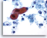
Figure 77
Sputum
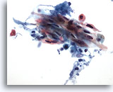
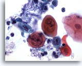
Atypical squamous metaplastic cells are considered suspicious for squamous cell carcinoma as their nuclei become hyperchromatic and angulated. 60X
Sputum
Atypical squamous metaplastic cells are considered suspicious for squamous cell carcinoma as their nuclei become hyperchromatic and angulated. 60X
Figure 77
Sputum
Atypical squamous metaplastic cells are considered suspicious for squamous cell carcinoma as their nuclei become hyperchromatic and angulated.
60X
Sputum
Atypical squamous metaplastic cells are considered suspicious for squamous cell carcinoma as their nuclei become hyperchromatic and angulated.
60X
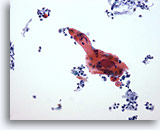
Figure 78
Bronchial wash
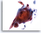
At lower magnification a large cell, highly suspicious for squamous cell carcinoma, is present. 20x
Bronchial wash
At lower magnification a large cell, highly suspicious for squamous cell carcinoma, is present. 20x
Figure 78
Bronchial wash
At lower magnification a large cell, highly suspicious for squamous cell carcinoma, is present.
20x
Bronchial wash
At lower magnification a large cell, highly suspicious for squamous cell carcinoma, is present.
20x
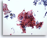
Figure 79
Bronchial wash
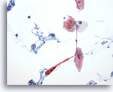
Numerous atypical squamous cells with polys are present in this cluster of cells from a case of squamous cell carcinoma. 20x
Bronchial wash
Numerous atypical squamous cells with polys are present in this cluster of cells from a case of squamous cell carcinoma. 20x
Figure 79
Bronchial wash
Numerous atypical squamous cells with polys are present in this cluster of cells from a case of squamous cell carcinoma.
20x
Bronchial wash
Numerous atypical squamous cells with polys are present in this cluster of cells from a case of squamous cell carcinoma.
20x
Figure 80
Bronchial wash
Spindle-shaped and filamentous cells from squamous cell carcinoma.
20x
Bronchial wash
Spindle-shaped and filamentous cells from squamous cell carcinoma.
20x
Figure 81
Bronchial wash
Higher magnification of spindle cells from squamous cell carcinoma
40x
Bronchial wash
Higher magnification of spindle cells from squamous cell carcinoma
40x
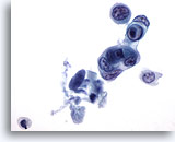
Figure 82
Bronchial wash
Not all cells of squamous cell carcinomas are orangeophilic. Basophilic staining may be seen. 60x
Bronchial wash
Not all cells of squamous cell carcinomas are orangeophilic. Basophilic staining may be seen. 60x
Figure 82
Bronchial wash
Not all cells of squamous cell carcinomas are orangeophilic. Basophilic staining may be seen.
60x
Bronchial wash
Not all cells of squamous cell carcinomas are orangeophilic. Basophilic staining may be seen.
60x
Figure 83
Bronchial wash
A pearl of keratinized cells from a case of squamous cell carcinoma.
60x
Bronchial wash
A pearl of keratinized cells from a case of squamous cell carcinoma.
60x
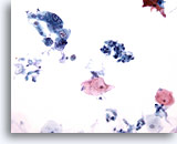
Figure 84
Bronchial wash
Lower magnification showing squamous cell carcinoma in the upper left portion of the field. 20x
Bronchial wash
Lower magnification showing squamous cell carcinoma in the upper left portion of the field. 20x
Figure 84
Bronchial wash
Lower magnification showing squamous cell carcinoma in the upper left portion of the field.
20x
Bronchial wash
Lower magnification showing squamous cell carcinoma in the upper left portion of the field.
20x
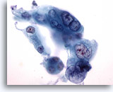
Figure 85
Bronchial wash
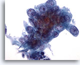
This cluster of cells from squamous cell carcinoma has marked nuclear atypia. The dense cytoplasm and sharp cytoplasmic border indicates squamous differentiation. 60x
Bronchial wash
This cluster of cells from squamous cell carcinoma has marked nuclear atypia. The dense cytoplasm and sharp cytoplasmic border indicates squamous differentiation. 60x
Figure 85
Bronchial wash
This cluster of cells from squamous cell carcinoma has marked nuclear atypia. The dense cytoplasm and sharp cytoplasmic border indicates squamous differentiation.
60x
Bronchial wash
This cluster of cells from squamous cell carcinoma has marked nuclear atypia. The dense cytoplasm and sharp cytoplasmic border indicates squamous differentiation.
60x
Figure 86
Bronchial wash
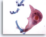
Tadpole shaped cells maybe seen in squamous cell carcinoma.
20x
Bronchial wash
Tadpole shaped cells maybe seen in squamous cell carcinoma.
20x
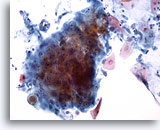
Figure 87
Bronchial wash
This cluster contains many abnormal cells. Their nuclear detail is seen at the periphery of the cluster. 20x
Bronchial wash
This cluster contains many abnormal cells. Their nuclear detail is seen at the periphery of the cluster. 20x
Figure 87
Bronchial wash
This cluster contains many abnormal cells. Their nuclear detail is seen at the periphery of the cluster.
20x
Bronchial wash
This cluster contains many abnormal cells. Their nuclear detail is seen at the periphery of the cluster.
20x
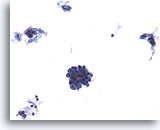
Figure 88
Bronchial wash
When in a cluster, it may be difficult to distinguish the cells of squamous cell carcinoma from adenocarcinoma. 20x
Bronchial wash
When in a cluster, it may be difficult to distinguish the cells of squamous cell carcinoma from adenocarcinoma. 20x
Figure 88
Bronchial wash
When in a cluster, it may be difficult to distinguish the cells of squamous cell carcinoma from adenocarcinoma.
20x
Bronchial wash
When in a cluster, it may be difficult to distinguish the cells of squamous cell carcinoma from adenocarcinoma.
20x
Figure 89
Bronchial wash
Poorly differentiated squamous cell carcinoma.
60x
Bronchial wash
Poorly differentiated squamous cell carcinoma.
60x
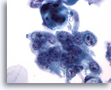
Figure 90
Bronchial wash
Cell borders in poorly-differentiated squamous cell carcinoma are sharper than those of adenocarcinoma. Nucleoli are multiple, and eccentric. 40x
Bronchial wash
Cell borders in poorly-differentiated squamous cell carcinoma are sharper than those of adenocarcinoma. Nucleoli are multiple, and eccentric. 40x
Figure 90
Bronchial wash
Cell borders in poorly-differentiated squamous cell carcinoma are sharper than those of adenocarcinoma. Nucleoli are multiple, and eccentric.
40x
Bronchial wash
Cell borders in poorly-differentiated squamous cell carcinoma are sharper than those of adenocarcinoma. Nucleoli are multiple, and eccentric.
40x
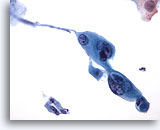
Figure 91
Bronchial wash
Poorly differentiated squamous cell carcinoma. Keratin material is present in vacuoles in the cytoplasm. 60x
Bronchial wash
Poorly differentiated squamous cell carcinoma. Keratin material is present in vacuoles in the cytoplasm. 60x
Figure 91
Bronchial wash
Poorly differentiated squamous cell carcinoma. Keratin material is present in vacuoles in the cytoplasm.
60x
Bronchial wash
Poorly differentiated squamous cell carcinoma. Keratin material is present in vacuoles in the cytoplasm.
60x
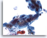
Figure 92
Bronchial wash
Careful study of a poorly differentiated squamous cell carcinoma shows scattered keratinization. 40x
Bronchial wash
Careful study of a poorly differentiated squamous cell carcinoma shows scattered keratinization. 40x
Figure 92
Bronchial wash
Careful study of a poorly differentiated squamous cell carcinoma shows scattered keratinization.
40x
Bronchial wash
Careful study of a poorly differentiated squamous cell carcinoma shows scattered keratinization.
40x
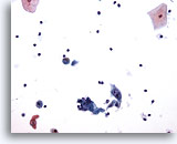
Figure 93
Bronchial wash
Low magnification shows atypical keratinized cells in squamous cell carcinoma. 20x
Bronchial wash
Low magnification shows atypical keratinized cells in squamous cell carcinoma. 20x
Figure 93
Bronchial wash
Low magnification shows atypical keratinized cells in squamous cell carcinoma.
20x
Bronchial wash
Low magnification shows atypical keratinized cells in squamous cell carcinoma.
20x
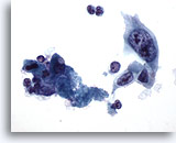
Figure 94
Bronchial wash
High magnification reveals bizarre nuclei, admixed with necrotic debris. 60x
Bronchial wash
High magnification reveals bizarre nuclei, admixed with necrotic debris. 60x
Figure 94
Bronchial wash
High magnification reveals bizarre nuclei, admixed with necrotic debris.
60x
Bronchial wash
High magnification reveals bizarre nuclei, admixed with necrotic debris.
60x
Figure 95
Bronchial wash
Deeply keratinized cells are seen in this squamous cell carcinoma.
60x
Bronchial wash
Deeply keratinized cells are seen in this squamous cell carcinoma.
60x
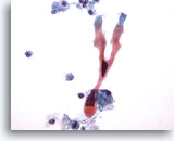
Figure 96
Bronchial wash
Bizarre cell shapes with sharply angulated nuclei in this squamous cell carcinoma. 60x
Bronchial wash
Bizarre cell shapes with sharply angulated nuclei in this squamous cell carcinoma. 60x
Figure 96
Bronchial wash
Bizarre cell shapes with sharply angulated nuclei in this squamous cell carcinoma.
60x
Bronchial wash
Bizarre cell shapes with sharply angulated nuclei in this squamous cell carcinoma.
60x
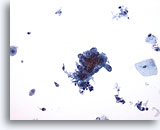
Figure 97
Bronchial brushing
Low magnification of a bronchial brushing of a squamous cell carcinoma. 20x
Bronchial brushing
Low magnification of a bronchial brushing of a squamous cell carcinoma. 20x
Figure 97
Bronchial brushing
Low magnification of a bronchial brushing of a squamous cell carcinoma.
20x
Bronchial brushing
Low magnification of a bronchial brushing of a squamous cell carcinoma.
20x
Figure 98
Bronchial brushing
The cells in this squamous cell carcinoma are well differentiated.
60x
Bronchial brushing
The cells in this squamous cell carcinoma are well differentiated.
60x
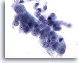
Figure 99
Bronchial brushing
Mildly reactive bronchial epithelium. Compare this to the previous photograph. 60x
Bronchial brushing
Mildly reactive bronchial epithelium. Compare this to the previous photograph. 60x
Figure 99
Bronchial brushing
Mildly reactive bronchial epithelium. Compare this to the previous photograph.
60x
Bronchial brushing
Mildly reactive bronchial epithelium. Compare this to the previous photograph.
60x
Figure 100
Bronchial brushing
Giant tumor cells are present in this squamous cell carcinoma
60x
Bronchial brushing
Giant tumor cells are present in this squamous cell carcinoma
60x
Figure 101
Left upper lobe brushing
Low magnification of a squamous cell carcinoma.
20x
Left upper lobe brushing
Low magnification of a squamous cell carcinoma.
20x
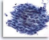
Figure 102
Left upper lobe brushing
Sheets of tumor cells are a feature of squamous cell carcinoma. 40x
Left upper lobe brushing
Sheets of tumor cells are a feature of squamous cell carcinoma. 40x
Figure 102
Left upper lobe brushing
Sheets of tumor cells are a feature of squamous cell carcinoma.
40x
Left upper lobe brushing
Sheets of tumor cells are a feature of squamous cell carcinoma.
40x
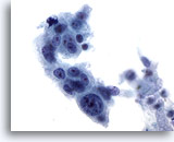
Figure 103
Left upper lobe brushing
Note in this case the variable number and position of nucleoli. 60x
Left upper lobe brushing
Note in this case the variable number and position of nucleoli. 60x
Figure 103
Left upper lobe brushing
Note in this case the variable number and position of nucleoli.
60x
Left upper lobe brushing
Note in this case the variable number and position of nucleoli.
60x
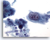
Figure 104
Left upper lobe brushing
The cell on the right shows irregular nuclear shape, clumped chromatin and multiple irregularly shaped nucleoli. There are masses of keratin in the cytoplasm indicating squamous differentiation. 60x
Left upper lobe brushing
The cell on the right shows irregular nuclear shape, clumped chromatin and multiple irregularly shaped nucleoli. There are masses of keratin in the cytoplasm indicating squamous differentiation. 60x
Figure 104
Left upper lobe brushing
The cell on the right shows irregular nuclear shape, clumped chromatin and multiple irregularly shaped nucleoli. There are masses of keratin in the cytoplasm indicating squamous differentiation.
60x
Left upper lobe brushing
The cell on the right shows irregular nuclear shape, clumped chromatin and multiple irregularly shaped nucleoli. There are masses of keratin in the cytoplasm indicating squamous differentiation.
60x

