Respiratory Cytology - Small Cell Undifferentiated Carcinoma
SMALL CELL UNDIFFERENTIATED CARCINOMA
Reminder: You may click on any slide image
for an enlarged view.
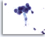
for an enlarged view.

Figure 142
Sputum
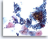
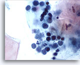
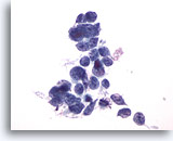
A sputum sample showing small cell undifferentiated carcinoma. Compare malignant cells’ size to the superficial squamous cells. 20x
Sputum
A sputum sample showing small cell undifferentiated carcinoma. Compare malignant cells’ size to the superficial squamous cells. 20x
Figure 142
Sputum
A sputum sample showing small cell undifferentiated carcinoma. Compare malignant cells’ size to the superficial squamous cells.
20x
Sputum
A sputum sample showing small cell undifferentiated carcinoma. Compare malignant cells’ size to the superficial squamous cells.
20x

Figure 143
Sputum
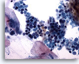
Nuclear staining ranges from dark to pale, reflecting pyknosis and cell necrosis, respectively. 40x
Sputum
Nuclear staining ranges from dark to pale, reflecting pyknosis and cell necrosis, respectively. 40x
Figure 143
Sputum
Nuclear staining ranges from dark to pale, reflecting pyknosis and cell necrosis, respectively.
40x
Sputum
Nuclear staining ranges from dark to pale, reflecting pyknosis and cell necrosis, respectively.
40x

Figure 144
Sputum
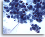
High magnification showing nuclear pyknosis in small cell undifferentiated carcinoma. 60x
Sputum
High magnification showing nuclear pyknosis in small cell undifferentiated carcinoma. 60x
Figure 144
Sputum
High magnification showing nuclear pyknosis in small cell undifferentiated carcinoma.
60x
Sputum
High magnification showing nuclear pyknosis in small cell undifferentiated carcinoma.
60x

Figure 145
Sputum
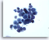
The molding of nuclei around each other, a feature of small cell undifferentiated carcinoma, is seen at the center of this group. 60x
Sputum
The molding of nuclei around each other, a feature of small cell undifferentiated carcinoma, is seen at the center of this group. 60x
Figure 145
Sputum
The molding of nuclei around each other, a feature of small cell undifferentiated carcinoma, is seen at the center of this group.
60x
Sputum
The molding of nuclei around each other, a feature of small cell undifferentiated carcinoma, is seen at the center of this group.
60x

Figure 146
Sputum
Nuclear pyknosis is more common in sputum samples of small cell undifferentiated carcinoma, compared to sample by direct collection. 60x
Sputum
Nuclear pyknosis is more common in sputum samples of small cell undifferentiated carcinoma, compared to sample by direct collection. 60x
Figure 146
Sputum
Nuclear pyknosis is more common in sputum samples of small cell undifferentiated carcinoma, compared to sample by direct collection.
60x
Sputum
Nuclear pyknosis is more common in sputum samples of small cell undifferentiated carcinoma, compared to sample by direct collection.
60x

Figure 147
Sputum
In sputum, the tumor cells of small cell undifferentiated carcinoma may be seen in a linear configuration. 60x
Sputum
In sputum, the tumor cells of small cell undifferentiated carcinoma may be seen in a linear configuration. 60x
Figure 147
Sputum
In sputum, the tumor cells of small cell undifferentiated carcinoma may be seen in a linear configuration.
60x
Sputum
In sputum, the tumor cells of small cell undifferentiated carcinoma may be seen in a linear configuration.
60x

Figure 148
Wang needle
This sample, collected by transbronchial needle biopsy, shows better nuclear preservation and less nuclear molding. 40x
Wang needle
This sample, collected by transbronchial needle biopsy, shows better nuclear preservation and less nuclear molding. 40x
Figure 148
Wang needle
This sample, collected by transbronchial needle biopsy, shows better nuclear preservation and less nuclear molding.
40x
Wang needle
This sample, collected by transbronchial needle biopsy, shows better nuclear preservation and less nuclear molding.
40x

Figure 149
Wang needle
Notice the delicate chromatin of this small cell undifferentiated carcinoma collected by needle aspiration. Nucleoli are not seen. 60x
Wang needle
Notice the delicate chromatin of this small cell undifferentiated carcinoma collected by needle aspiration. Nucleoli are not seen. 60x
Figure 149
Wang needle
Notice the delicate chromatin of this small cell undifferentiated carcinoma collected by needle aspiration. Nucleoli are not seen.
60x
Wang needle
Notice the delicate chromatin of this small cell undifferentiated carcinoma collected by needle aspiration. Nucleoli are not seen.
60x

Figure 150
Wang needle
Cells may be rounded, carrot shaped or pyramidal in small cell undifferentiated carcinoma. 60x
Wang needle
Cells may be rounded, carrot shaped or pyramidal in small cell undifferentiated carcinoma. 60x
Figure 150
Wang needle
Cells may be rounded, carrot shaped or pyramidal in small cell undifferentiated carcinoma.
60x
Wang needle
Cells may be rounded, carrot shaped or pyramidal in small cell undifferentiated carcinoma.
60x

Figure 151
Bronchial brushing
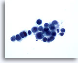
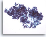
Bronchial brushing of small cell undifferentiated carcinoma. Cells are tightly clustered. 40x
Bronchial brushing
Bronchial brushing of small cell undifferentiated carcinoma. Cells are tightly clustered. 40x
Figure 151
Bronchial brushing
Bronchial brushing of small cell undifferentiated carcinoma. Cells are tightly clustered.
40x
Bronchial brushing
Bronchial brushing of small cell undifferentiated carcinoma. Cells are tightly clustered.
40x

Figure 152
Bronchial brushing
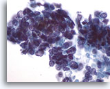
Higher magnification shows nuclear molding and individual cell necrosis. 60x
Bronchial brushing
Higher magnification shows nuclear molding and individual cell necrosis. 60x
Figure 152
Bronchial brushing
Higher magnification shows nuclear molding and individual cell necrosis.
60x
Bronchial brushing
Higher magnification shows nuclear molding and individual cell necrosis.
60x

Figure 153
Bronchoalveolar lavage
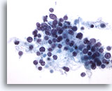
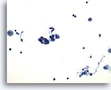
The intermediate cell type of small cell undifferentiated carcinoma is slightly larger in cell diameter. 20x
Bronchoalveolar lavage
The intermediate cell type of small cell undifferentiated carcinoma is slightly larger in cell diameter. 20x
Figure 153
Bronchoalveolar lavage
The intermediate cell type of small cell undifferentiated carcinoma is slightly larger in cell diameter.
20x
Bronchoalveolar lavage
The intermediate cell type of small cell undifferentiated carcinoma is slightly larger in cell diameter.
20x

Figure 154
Bronchoalveolar lavage
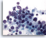
Small cell undifferentiated carcinoma, intermediate cell type. Compare the malignant cells’ size to the adjacent columnar bronchial epithelial cell. 60x
Bronchoalveolar lavage
Small cell undifferentiated carcinoma, intermediate cell type. Compare the malignant cells’ size to the adjacent columnar bronchial epithelial cell. 60x
Figure 154
Bronchoalveolar lavage
Small cell undifferentiated carcinoma, intermediate cell type. Compare the malignant cells’ size to the adjacent columnar bronchial epithelial cell.
60x
Bronchoalveolar lavage
Small cell undifferentiated carcinoma, intermediate cell type. Compare the malignant cells’ size to the adjacent columnar bronchial epithelial cell.
60x
