Respiratory Cytology - Reactive Inflammatory
REACTIVE INFLAMMATORY
Reminder: You may click on any slide image
for an enlarged view.
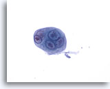
Figure 22
Bronchial wash
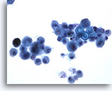
Multinucleated epithelial cells with intranuclear inclusions are characteristic of Herpes simplex infection of the lung. 60x
Bronchial wash
Multinucleated epithelial cells with intranuclear inclusions are characteristic of Herpes simplex infection of the lung. 60x
Figure 22
Bronchial wash
Multinucleated epithelial cells with intranuclear inclusions are characteristic of Herpes simplex infection of the lung.
60x
Bronchial wash
Multinucleated epithelial cells with intranuclear inclusions are characteristic of Herpes simplex infection of the lung.
60x
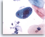
Figure 23
Bronchial wash
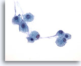
Compare the size of this multinucleated cell infected by Herpes to the adjacent superficial squamous cells. 60x
Bronchial wash
Compare the size of this multinucleated cell infected by Herpes to the adjacent superficial squamous cells. 60x
Figure 23
Bronchial wash
Compare the size of this multinucleated cell infected by Herpes to the adjacent superficial squamous cells.
60x
Bronchial wash
Compare the size of this multinucleated cell infected by Herpes to the adjacent superficial squamous cells.
60x
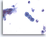
Figure 24
Bronchial wash
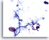
Herpes simplex infection may cause single intranuclear inclusions as illustrated in these epithelial cells. 60x
Bronchial wash
Herpes simplex infection may cause single intranuclear inclusions as illustrated in these epithelial cells. 60x
Figure 24
Bronchial wash
Herpes simplex infection may cause single intranuclear inclusions as illustrated in these epithelial cells.
60x
Bronchial wash
Herpes simplex infection may cause single intranuclear inclusions as illustrated in these epithelial cells.
60x
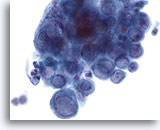
Figure 25
Bronchial wash
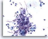
The inclusions of H. simplex can also cause a “glassy” appearance to the nucleus. 60x
Bronchial wash
The inclusions of H. simplex can also cause a “glassy” appearance to the nucleus. 60x
Figure 25
Bronchial wash
The inclusions of H. simplex can also cause a “glassy” appearance to the nucleus.
60x
Bronchial wash
The inclusions of H. simplex can also cause a “glassy” appearance to the nucleus.
60x
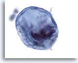
Figure 26
Bronchial wash
Vegetable cells in respiratory samples may mimic atypical epithelial cells or cancer. 60x
Bronchial wash
Vegetable cells in respiratory samples may mimic atypical epithelial cells or cancer. 60x
Figure 26
Bronchial wash
Vegetable cells in respiratory samples may mimic atypical epithelial cells or cancer.
60x
Bronchial wash
Vegetable cells in respiratory samples may mimic atypical epithelial cells or cancer.
60x
Figure 27
Bronchial wash
Clusters of actinomycetes commonly contaminate respiratory samples
60x
Bronchial wash
Clusters of actinomycetes commonly contaminate respiratory samples
60x
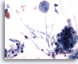
Figure 28
Bronchial wash
Hyphae of Candida species may represent oral contamination of the sample or underlying fungal pneumonia. 40x
Bronchial wash
Hyphae of Candida species may represent oral contamination of the sample or underlying fungal pneumonia. 40x
Figure 28
Bronchial wash
Hyphae of Candida species may represent oral contamination of the sample or underlying fungal pneumonia.
40x
Bronchial wash
Hyphae of Candida species may represent oral contamination of the sample or underlying fungal pneumonia.
40x
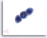
Figure 29
Bronchial wash
Atypical metaplastic cells can accompany fungal infections of the lung. This finding is particularly striking with aspergillus infection. 60x
Bronchial wash
Atypical metaplastic cells can accompany fungal infections of the lung. This finding is particularly striking with aspergillus infection. 60x
Figure 29
Bronchial wash
Atypical metaplastic cells can accompany fungal infections of the lung. This finding is particularly striking with Aspergillus infection. 60x
Bronchial wash
Atypical metaplastic cells can accompany fungal infections of the lung. This finding is particularly striking with Aspergillus infection. 60x
Figure 30
Bronchial wash
Bronchial washing showing zygomycetes in an inflammatory background.
10x
Bronchial wash
Bronchial washing showing zygomycetes in an inflammatory background.
10x
Figure 31
Bronchial wash
Even with just Pap stain, zygomycetes have brown-pigmented hyphae. 20x
Bronchial wash
Even with just Pap stain, zygomycetes have brown-pigmented hyphae. 20x
Figure 32
Bronchial wash
Hyphal forms of zygomycetes in a bronchial wash specimen.
40x
Bronchial wash
Hyphal forms of zygomycetes in a bronchial wash specimen.
40x
Figure 33
Bronchial wash
The ribbon-like nature of these wide hyphae is shown.
40x
Bronchial wash
The ribbon-like nature of these wide hyphae is shown.
40x
Figure 34
Lung wash
Aspergillus spp. is characterized by septate hyphae with parallel walls.
20x
Lung wash
Aspergillus spp. is characterized by septate hyphae with parallel walls.
20x
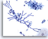
Figure 35
Bronchial wash
This cluster of aspergillus illustrates the 45 degree branching that is typical of the organism. 40x
Bronchial wash
This cluster of aspergillus illustrates the 45 degree branching that is typical of the organism. 40x
Figure 35
Bronchial wash
This cluster of Aspergillus illustrates the 45 degree branching that is typical of the organism.
40x
Bronchial wash
This cluster of Aspergillus illustrates the 45 degree branching that is typical of the organism.
40x
Figure 36
Bronchial wash
Screening magnification showing Aspergillus spp. 10x
Bronchial wash
Screening magnification showing Aspergillus spp. 10x
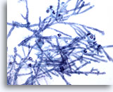
Figure 37
Bronchial wash
Higher power magnification shows the parallel cell walls with septate hyphae. 40x
Bronchial wash
Higher power magnification shows the parallel cell walls with septate hyphae. 40x
Figure 37
Bronchial wash
Higher power magnification shows the parallel cell walls with septate hyphae.
40x
Bronchial wash
Higher power magnification shows the parallel cell walls with septate hyphae.
40x
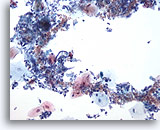
Figure 38
Bronchial wash
Yeast-like fungi may be seen admixed with oral squamous cells. These most likely represent contamination. 20x
Bronchial wash
Yeast-like fungi may be seen admixed with oral squamous cells. These most likely represent contamination. 20x
Figure 38
Bronchial wash
Yeast-like fungi may be seen admixed with oral squamous cells. These most likely represent contamination.
20x
Bronchial wash
Yeast-like fungi may be seen admixed with oral squamous cells. These most likely represent contamination.
20x
Figure 39
Bronchial wash
Numerous small yeast are seen in this photomicrograph.
60x
Bronchial wash
Numerous small yeast are seen in this photomicrograph.
60x
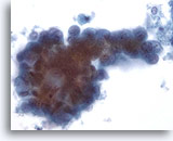
Figure 40
Bronchial wash
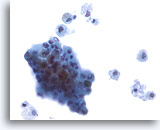
Clusters of reactive bronchial epithelial cells a commonly accompany aspergillus infections. 60x
Bronchial wash
Clusters of reactive bronchial epithelial cells a commonly accompany aspergillus infections. 60x
Figure 40
Bronchial wash
Clusters of reactive bronchial epithelial cells a commonly accompany Aspergillus infections.
60x
Bronchial wash
Clusters of reactive bronchial epithelial cells a commonly accompany Aspergillus infections.
60x
Figure 41
Bronchial wash
Atypical metaplastic cells may be seen with Aspergillus infection.
60x
Bronchial wash
Atypical metaplastic cells may be seen with Aspergillus infection.
60x
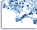
Figure 42
Bronchial wash
A reactive multinucleated bronchial epithelial cell is present in a background of necrosis. 60x
Bronchial wash
A reactive multinucleated bronchial epithelial cell is present in a background of necrosis. 60x
Figure 42
Bronchial wash
A reactive multinucleated bronchial epithelial cell is present in a background of necrosis.
60x
Bronchial wash
A reactive multinucleated bronchial epithelial cell is present in a background of necrosis.
60x
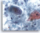
Figure 43
Bronchial wash
The necrotic material associated with an aspergilloma may harbor reactive cells. 60x
Bronchial wash
The necrotic material associated with an aspergilloma may harbor reactive cells. 60x
Figure 43
Bronchial wash
The necrotic material associated with an aspergilloma may harbor reactive cells.
60x
Bronchial wash
The necrotic material associated with an aspergilloma may harbor reactive cells.
60x
Figure 44
Bronchial wash
High magnification showing septate hyphae of Aspergillus.
40x
Bronchial wash
High magnification showing septate hyphae of Aspergillus.
40x
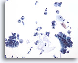
Figure 45
Bronchial wash
Asthma or chronic bronchitis may cause hyperplasia of bronchial epithelial cells. 20x
Bronchial wash
Asthma or chronic bronchitis may cause hyperplasia of bronchial epithelial cells. 20x
Figure 45
Bronchial wash
Asthma or chronic bronchitis may cause hyperplasia of bronchial epithelial cells.
20x
Bronchial wash
Asthma or chronic bronchitis may cause hyperplasia of bronchial epithelial cells.
20x
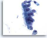
Figure 46
Bronchial wash
Hyperplastic bronchial epithelial cells are enlarged, with nuclei that may be multiple times the diameter of normal. 60x
Bronchial wash
Hyperplastic bronchial epithelial cells are enlarged, with nuclei that may be multiple times the diameter of normal. 60x
Figure 46
Bronchial wash
Hyperplastic bronchial epithelial cells are enlarged, with nuclei that may be multiple times the diameter of normal.
60x
Bronchial wash
Hyperplastic bronchial epithelial cells are enlarged, with nuclei that may be multiple times the diameter of normal.
60x
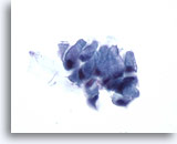
Figure 47
Bronchial wash
Goblet cells are seen in conjunction with chronic bronchitis and asthma. 60x
Bronchial wash
Goblet cells are seen in conjunction with chronic bronchitis and asthma. 60x
Figure 47
Bronchial wash
Goblet cells are seen in conjunction with chronic bronchitis and asthma.
60x
Bronchial wash
Goblet cells are seen in conjunction with chronic bronchitis and asthma.
60x
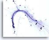
Figure 48
Bronchial wash
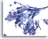
Curschmann’s spirals represent inspissated mucus that forms as a cast of a bronchiole. 20x
Bronchial wash
Curschmann’s spirals represent inspissated mucus that forms as a cast of a bronchiole. 20x
Figure 48
Bronchial wash
Curschmann’s spirals represent inspissated mucus that forms as a cast of a bronchiole.
20x
Bronchial wash
Curschmann’s spirals represent inspissated mucus that forms as a cast of a bronchiole.
20x
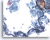
Figure 49
Bronchial wash
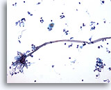
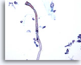
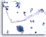
Fiber material contaminating the lung may resemble a Curschmann’s spiral. 20x
Bronchial wash
Fiber material contaminating the lung may resemble a Curschmann’s spiral. 20x
Figure 49
Bronchial wash
Fiber material contaminating the lung may resemble a Curschmann’s spiral.
20x
Bronchial wash
Fiber material contaminating the lung may resemble a Curschmann’s spiral.
20x
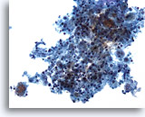
Figure 50
Bronchoalveolar lavage
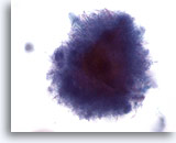
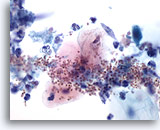
Bronchoalvelar lavage can be used to sample peripheral airways for infections. A cluster of Pneumocystis carinii is shown in the lower left. 20x
Bronchoalveolar lavage
Bronchoalvelar lavage can be used to sample peripheral airways for infections. A cluster of Pneumocystis carinii is shown in the lower left. 20x
Figure 50
Bronchoalveolar lavage
Bronchoalvelar lavage can be used to sample peripheral airways for infections. A cluster of Pneumocystis carinii is shown in the lower left.
20x
Bronchoalveolar lavage
Bronchoalvelar lavage can be used to sample peripheral airways for infections. A cluster of Pneumocystis carinii is shown in the lower left.
20x
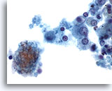
Figure 51
Bronchoalveolar lavage
Higher magnification of P. carinii adjacent to respiratory debris. 60x
Bronchoalveolar lavage
Higher magnification of P. carinii adjacent to respiratory debris. 60x
Figure 51
Bronchoalveolar lavage
Higher magnification of P. carinii adjacent to respiratory debris.
60x
Bronchoalveolar lavage
Higher magnification of P. carinii adjacent to respiratory debris.
60x
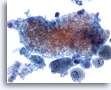
Figure 52
Bronchoalveolar lavage
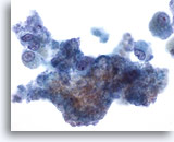
The cyst forms of P. carinii form a foamy mass within the sample. 60x
Bronchoalveolar lavage
The cyst forms of P. carinii form a foamy mass within the sample. 60x
Figure 52
Bronchoalveolar lavage
The cyst forms of P. carinii form a foamy mass within the sample.
60x
Bronchoalveolar lavage
The cyst forms of P. carinii form a foamy mass within the sample.
60x
Figure 53
Bronchoalveolar lavage
Another example of foamy clusters of P. carinii. 60x
Bronchoalveolar lavage
Another example of foamy clusters of P. carinii. 60x
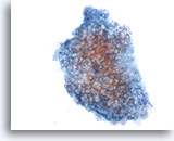
Figure 54
Bronchoalveolar lavage
The foamy masses of P. carinii are literally casts of the alveoli. 60x
Bronchoalveolar lavage
The foamy masses of P. carinii are literally casts of the alveoli. 60x
Figure 54
Bronchoalveolar lavage
The foamy masses of P. carinii are literally casts of the alveoli.
60x
Bronchoalveolar lavage
The foamy masses of P. carinii are literally casts of the alveoli.
60x
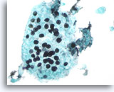
Figure 55
Bronchoalveolar lavage
Methenamine silver stain illustrates the trophozoites of P. carinii. 60x
Bronchoalveolar lavage
Methenamine silver stain illustrates the trophozoites of P. carinii. 60x
Figure 55
Bronchoalveolar lavage
Methenamine silver stain illustrates the trophozoites of P. carinii.
60x
Bronchoalveolar lavage
Methenamine silver stain illustrates the trophozoites of P. carinii.
60x
Figure 56
Bronchoalveolar lavage
Bronchoalveolar lavage showing metaplastic squamous cells.
40x
Bronchoalveolar lavage
Bronchoalveolar lavage showing metaplastic squamous cells.
40x
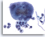
Figure 57
Bronchoalveolar lavage
This multinucleated giant histiocyte could be mistaken for a reactive cluster of bronchial epithelial cells. Nuclei retain benign morphology. 60x
Bronchoalveolar lavage
This multinucleated giant histiocyte could be mistaken for a reactive cluster of bronchial epithelial cells. Nuclei retain benign morphology. 60x
Figure 57
Bronchoalveolar lavage
This multinucleated giant histiocyte could be mistaken for a reactive cluster of bronchial epithelial cells. Nuclei retain benign morphology.
60x
Bronchoalveolar lavage
This multinucleated giant histiocyte could be mistaken for a reactive cluster of bronchial epithelial cells. Nuclei retain benign morphology.
60x
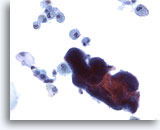
Figure 58
Bronchoalveolar lavage
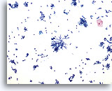
Oral-derived bacteria may be found in the bronchoalveolar lavage sample. 40x
Bronchoalveolar lavage
Oral-derived bacteria may be found in the bronchoalveolar lavage sample. 40x
Figure 58
Bronchoalveolar lavage
Oral-derived bacteria may be found in the bronchoalveolar lavage sample.
40x
Bronchoalveolar lavage
Oral-derived bacteria may be found in the bronchoalveolar lavage sample.
40x
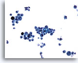
Figure 59
Bronchoalveolar lavage
Low power magnification showing Blastomyces among respiratory macrophages. 20x
Bronchoalveolar lavage
Low power magnification showing Blastomyces among respiratory macrophages. 20x
Figure 59
Bronchoalveolar lavage
Low power magnification showing Blastomyces among respiratory macrophages.
20x
Bronchoalveolar lavage
Low power magnification showing Blastomyces among respiratory macrophages.
20x
Figure 60
Bronchoalveolar lavage
Higher magnification shows large, thick walled fungi.
40x
Bronchoalveolar lavage
Higher magnification shows large, thick walled fungi.
40x
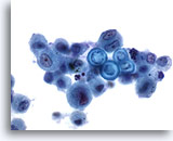
Figure 61
Bronchoalveolar lavage
Broad-based budding and a thick refractile wall characterize Blastomycoses. 60x
Bronchoalveolar lavage
Broad-based budding and a thick refractile wall characterize Blastomycoses. 60x
Figure 61
Bronchoalveolar lavage
Broad-based budding and a thick refractile wall characterize Blastomycoses.
60x
Bronchoalveolar lavage
Broad-based budding and a thick refractile wall characterize Blastomycoses.
60x
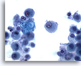
Figure 62
Bronchoalveolar lavage
Compare the size of B. dermatitidis with the nearby macrophages. 60x
Bronchoalveolar lavage
Compare the size of B. dermatitidis with the nearby macrophages. 60x
Figure 62
Bronchoalveolar lavage
Compare the size of B. dermatitidis with the nearby macrophages.
60x
Bronchoalveolar lavage
Compare the size of B. dermatitidis with the nearby macrophages.
60x
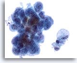
Figure 63
Bronchoalveolar lavage
Bronchoalveolar lavage collects macrophages, respiratory epithelia and other cells from the airways. BAL is commonly used to evaluate inflammatory and infectious disorders of the lung. 60x
Bronchoalveolar lavage
Bronchoalveolar lavage collects macrophages, respiratory epithelia and other cells from the airways. BAL is commonly used to evaluate inflammatory and infectious disorders of the lung. 60x
Figure 63
Bronchoalveolar lavage
Bronchoalveolar lavage collects macrophages, respiratory epithelia and other cells from the airways. BAL is commonly used to evaluate inflammatory and infectious disorders of the lung.
60x
Bronchoalveolar lavage
Bronchoalveolar lavage collects macrophages, respiratory epithelia and other cells from the airways. BAL is commonly used to evaluate inflammatory and infectious disorders of the lung.
60x
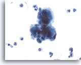
Figure 64
Bronchoalveolar lavage
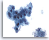
A benign cluster of epithelial cells is illustrated from this bronchoalveolar lavage sample. 40x
Bronchoalveolar lavage
A benign cluster of epithelial cells is illustrated from this bronchoalveolar lavage sample. 40x
Figure 64
Bronchoalveolar lavage
A benign cluster of epithelial cells is illustrated from this bronchoalveolar lavage sample.
40x
Bronchoalveolar lavage
A benign cluster of epithelial cells is illustrated from this bronchoalveolar lavage sample.
40x
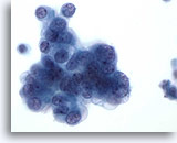
Figure 65
Bronchoalveolar lavage
Benign cells of terminal bronchiolar origin are shown. Note the vacuolated cytoplasm. 40x
Bronchoalveolar lavage
Benign cells of terminal bronchiolar origin are shown. Note the vacuolated cytoplasm. 40x
Figure 65
Bronchoalveolar lavage
Benign cells of terminal bronchiolar origin are shown. Note the vacuolated cytoplasm.
40x
Bronchoalveolar lavage
Benign cells of terminal bronchiolar origin are shown. Note the vacuolated cytoplasm.
40x
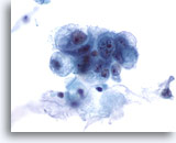
Figure 66
Bronchoalveolar lavage
Reactive/atypical cells of terminal bronchiolar/alveolar origin. Note the single, central nucleolus. 60x
Bronchoalveolar lavage
Reactive/atypical cells of terminal bronchiolar/alveolar origin. Note the single, central nucleolus. 60x
Figure 66
Bronchoalveolar lavage
Reactive/atypical cells of terminal bronchiolar/alveolar origin. Note the single, central nucleolus.
60x
Bronchoalveolar lavage
Reactive/atypical cells of terminal bronchiolar/alveolar origin. Note the single, central nucleolus.
60x
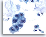
Figure 67
Bronchoalveolar lavage
Reactive/atypical terminal bronchiolar cells from a bronchoalveolar lavage. 60X
Bronchoalveolar lavage
Reactive/atypical terminal bronchiolar cells from a bronchoalveolar lavage. 60X
Figure 67
Bronchoalveolar lavage
Reactive/atypical terminal bronchiolar cells from a bronchoalveolar lavage.
60X
Bronchoalveolar lavage
Reactive/atypical terminal bronchiolar cells from a bronchoalveolar lavage.
60X
Figure 68
Sputum
This sputum sample contains respiratory macrophages, a criterion of adequacy.
40x
Sputum
This sputum sample contains respiratory macrophages, a criterion of adequacy.
40x
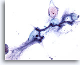
Figure 69
Sputum
A Curschmann’s spiral, a cast of a small bronchiole, in a sputum sample, as may be seen in asthma. 20x
Sputum
A Curschmann’s spiral, a cast of a small bronchiole, in a sputum sample, as may be seen in asthma. 20x
Figure 69
Sputum
A Curschmann’s spiral, a cast of a small bronchiole, in a sputum sample, as may be seen in asthma.
20x
Sputum
A Curschmann’s spiral, a cast of a small bronchiole, in a sputum sample, as may be seen in asthma.
20x
Figure 70
Sputum
Another example of a Curschmann’s spiral.
20x
Sputum
Another example of a Curschmann’s spiral.
20x
Figure 71
Sputum
Candida spp. , from the mouth, commonly contaminate sputum samples.
40x
Sputum
Candida spp. , from the mouth, commonly contaminate sputum samples.
40x
Figure 72
Actinomycetes
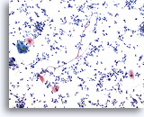
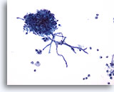
Actinomycetes may be present in sputum. Note the filamentous bacteria.
60x
Actinomycetes
Actinomycetes may be present in sputum. Note the filamentous bacteria.
60x
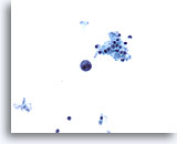
Figure 73
Bronchial wash
Low power magnification of cytomegalovirus infected cells in a bronchial wash sample. 20x
Bronchial wash
Low power magnification of cytomegalovirus infected cells in a bronchial wash sample. 20x
Figure 73
Bronchial wash
Low power magnification of cytomegalovirus infected cells in a bronchial wash sample.
20x
Bronchial wash
Low power magnification of cytomegalovirus infected cells in a bronchial wash sample.
20x
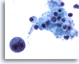
Figure 74
Bronchial wash
Higher magnification shows a bi-nucleated macrophage with intranuclear inclusions of cytomegalovirus. 60x
Bronchial wash
Higher magnification shows a bi-nucleated macrophage with intranuclear inclusions of cytomegalovirus. 60x
Figure 74
Bronchial wash
Higher magnification shows a bi-nucleated macrophage with intranuclear inclusions of cytomegalovirus.
60x
Bronchial wash
Higher magnification shows a bi-nucleated macrophage with intranuclear inclusions of cytomegalovirus.
60x
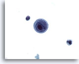
Figure 75
Bronchial wash
A single macrophage with both intranuclear and intracytoplasmic inclusions of CMV. 60x
Bronchial wash
A single macrophage with both intranuclear and intracytoplasmic inclusions of CMV. 60x
Figure 75
Bronchial wash
A single macrophage with both intranuclear and intracytoplasmic inclusions of CMV.
60x
Bronchial wash
A single macrophage with both intranuclear and intracytoplasmic inclusions of CMV.
60x

