Cytology of Pleural, Pericardial and Peritoneal Cavity Effusions - Effusions and Washes Ovarian Adenocarcinoma
EFFUSIONS AND WASHES – OVARIAN ADENOCARCINOMA
An unexplained peritoneal effusion in a female patient should prompt an assessment for malignant cells of ovarian origin
Reminder: You may click on any slide image
for an enlarged view.
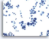
Pleural effusion:
Papillary serous ovarian adenocarcinoma.
20X
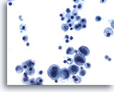
Pleural effusion:
Papillary serous ovarian adenocarcinoma.
40X
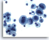
Pleural effusion:
Papillary serous ovarian adenocarcinoma.
60X
Figures 43-45: Pleural effusion Papillary serous ovarian adenocarcinoma.
Cells of papillary serous ovarian adenocarcinoma in a pleural effusion represent a discontinuous population of cells. Their cell and nuclear size is variable. Increased nuclear to cytoplasmic ratio and cytoplasmic vacuoles are features. Cells may exist singly or in small acinar groups.
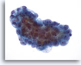
Pleural effusion: Serous ovarian adenocarcinoma. These tumor cells, metastatic from an ovarian adenocarcinoma and found in a pleural effusion, were shed as a three-dimensional papillary – glandular structure. 40X
Pleural effusion:
Serous ovarian adenocarcinoma. These tumor cells, metastatic from an ovarian adenocarcinoma and found in a pleural effusion, were shed as a three-dimensional papillary – glandular structure.
40X
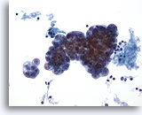
Pleural effusion: Serous ovarian adenocarcinoma. Though three-dimensional, the malignant nature of these ovarian cancer cells is easily appreciated when comparing their size to lymphocytes in the background. Distinction between ovarian and endometrial sources may be difficult given the common Mullerian origin of tumors in each site. 20X
Pleural effusion:
Serous ovarian adenocarcinoma. Though three-dimensional, the malignant nature of these ovarian cancer cells is easily appreciated when comparing their size to lymphocytes in the background. Distinction between ovarian and endometrial sources may be difficult given the common Mullerian origin of tumors in each site.
20X
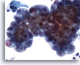
Pleural effusion: Serous ovarian adenocarcinoma. Sheets of tumor cells with large nuclei and prominent nucleoli. Again note the size difference in comparison to background lymphocytes. This is a discontinuous cell population. 40X
Pleural effusion:
Serous ovarian adenocarcinoma. Sheets of tumor cells with large nuclei and prominent nucleoli. Again note the size difference in comparison to background lymphocytes. This is a discontinuous cell population.
40X
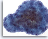
Peritoneal effusion: Serous borderline tumor. Papillary cluster of glandular serous borderline tumor forming rounded “cannon-balls” of tumor cells. Note the smooth border, compared to the scalloped border of reactive mesothelial cells 60X
Peritoneal effusion:
Serous borderline tumor. Papillary cluster of glandular serous borderline tumor forming rounded “cannon-balls” of tumor cells. Note the smooth border, compared to the scalloped border of reactive mesothelial cells
60X
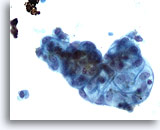
Peritoneal wash: Ovarian serous borderline tumor. Papillary cluster of tumor cells with prominent cytoplasmic vacuolization. 40X
Peritoneal wash:
Ovarian serous borderline tumor. Papillary cluster of tumor cells with prominent cytoplasmic vacuolization.
40X
Vigorous peritoneal washes may dislodge microscopic tumor. Washes are an integral part of staging laparoscopy. Because of the washing procedure, tumor cells generally come off in three- dimensional cohesive groups and may be admixed with sheets of benign mesothelium. The tumor cells are easily distinguished by size, malignant characteristics and crowded configurations.
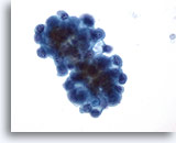
Pelvic wash: Ovarian serous borderline tumor. This pelvic wash has produced a three-dimensional crowded cluster of tumor cells with minimal atypical nuclear features. 40X
Pelvic wash:
Ovarian serous borderline tumor. This pelvic wash has produced a three-dimensional crowded cluster of tumor cells with minimal atypical nuclear features.
40X
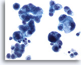
Peritoneal effusion: Serous ovarian adenocarcinoma. Pleomorphic, papillary groups of tumor cells, found in a peritoneal effusion. Note the size difference between tumor cells and the benign mesothelial cells at lower right. 40X
Peritoneal effusion:
Serous ovarian adenocarcinoma. Pleomorphic, papillary groups of tumor cells, found in a peritoneal effusion. Note the size difference between tumor cells and the benign mesothelial cells at lower right.
40X
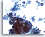
Peritoneal effusion: Ovarian adenocarcinoma. A three-dimensional tumor cell group is seen along with a “dirty” background. 20X
Peritoneal effusion:
Ovarian adenocarcinoma. A three-dimensional tumor cell group is seen along with a “dirty” background.
20X
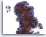
Pelvic wash: Ovarian adenocarcinoma. A malignant three-dimensional cell group contrasts with a group of four benign cells at upper left. 40X
Pelvic wash:
Ovarian adenocarcinoma. A malignant three-dimensional cell group contrasts with a group of four benign cells at upper left.
40X

